
Approaching the topic
- Understanding the key concepts
- Application of these concepts
Basic terms to ponder
- Genetics: The study of genes, variation and heredity.
- Locus: A position on chromosome where the gene is present.
- Genotype: The set of representing genes or alleles.
- Phenotype: This represents the characteristics (trait) expressed.
- Co-dominance: The condition in which neither of the alleles is dominant or recessive.
Genes
Location and function
- A heritable factor that consists of a DNA sequence and influences a specific character or trait in an individual.
- These are present on the locus of the chromosome. There can be many loci on a single chromosome which possesses different genes responsible for translating into different proteins.
Alleles
- These are the versions or a form of gene, differs with only one or few bases.
- Difference between the two alleles of the same gene can be seen in genetic diseases like complete achromatopsia and cystic fibrosis.
Transducin protein is necessary for the coloured vision from the eye. In this disease, the gene that controls the production of transducin protein, found on chromosome 1 becomes damaged or altered by mutation. The base sequence of this gene gets altered at 229th position with ‘C’ gets replaced by ‘T’ in one of the two contrasting pair of alleles. This makes two different alleles of the same gene. One is the standard allele with no defects and the other is rare mutated allele.
This disease is caused when the gene on chromosome 7 named CFTR, got altered. CFTR is responsible for maintaining the production of mucus in many parts of the body. This genetic disorder causes production of abnormally excessive quantities of mucus in various organs. The standard allele of the gene is unaltered and causes no defect but the mutated allele of CFTR gene causes cystic fibrosis.
- The production of new allele is caused by mutation.
Mutation
- It is a sudden, random change in the genetic material.
- Most common and significant is base substitution mutation, which results in a single base being changed by replacing it with another.
- For example;
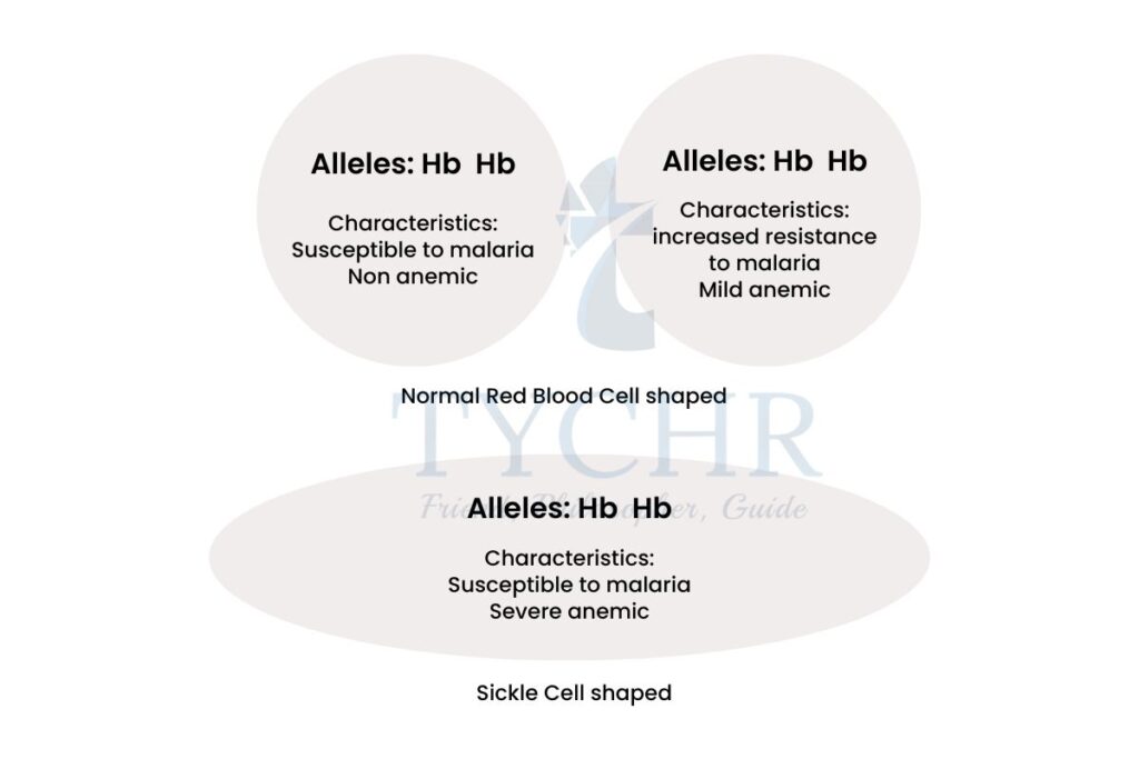
- Sickle cell disease:
- In this disease, the red blood cells changed their shape from biconcave to sickle shaped. It reduces their oxygen carrying capacity and causes severe anaemia and therefore it is also known as sickle cell anaemia.
- It occurs due to substitution of the base at the 6th codon of the haemoglobin chain. The codon GAG (codes for glutamate) becomes GTG (valine).
- Valine has different properties and effects than from glutamate therefore it produces complications within haemoglobin molecule.
- The normal allele of haemoglobin is HbA and the mutated allele causing this disease is HbS.
- Since the oxygen carrying capacity of the haemoglobin molecule becomes very less, so the symptoms shown by the individual with this disease are weakness, fatigue, shortness of breath etc.
- Can Sickle cell anaemia be good for us in any aspect?
- Yes. The person suffering from this disease will be resistant to malaria infection. Malaria is caused by a malarial parasite Plasmodium, which is transmitted to human blood by female Anopheles mosquito. Plasmodium attacks red blood cells and causes high fever and chills. The sickle shaped cells reduce the chances of survival of malarial parasite in the blood due to chemical imbalance (insufficient quantities of potassium).
- This is boon for those who inherit one copy of sickle trait and one copy of standard disc-shaped cells they will be having some disc shaped and some sickle shaped cells and are not affected by anaemia.
- Can mutation be good for us in any aspect?
- Yes. Mutation in LRP5 gene can help to prevent the attack of HIV (human immunodeficiency virus). LRP5 gene helps the immune system cells to make certain type of protein which acts as a receptor for HIV, to infect the other cells. So, if there is a mutated allele in the LRP5 gene then, HIV won’t affect the individual.
- Sickle cell disease:
Exam tip! While answering questions about “mutation”, elucidate your answer by giving examples of genetic diseases related to mutation. Drawing interlinkages with Human Genome Project will ensure maximum possible credits. |
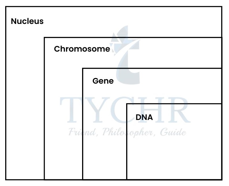
Genome
The complete set of organism’s base sequences of DNA is called its genome.
- A short DNA fragment (containing 21 bases) looks like GTG CAC CTG ACT CCT GAG GAG, each letter representing one of the four nucleotide bases.
- Whole genome of humans is 46 chromosomes in the nucleus + DNA of mitochondrion, while in plants it is chromosomes in the nucleus + DNA of mitochondrion and chloroplast.
- The Human Genome Project was begun in 1990, with the aim to find all the base sequences of the human genome. The conclusions and features of Human Genome Project are:
- There are approximately 23,000 genes present in the whole human genome.
- Most of the genome was not transcribed.
- Thousands of loci are discovered among 23 pairs of chromosomes.
- There are 3164.7 million bp in human genome, with average gene consisting of 3,000 bases.
- 99.9 percent of nucleotide bases are exactly same in every human i.e. only 0.1 percent of the sequence makes us different.
- Less than 2% of the genome codes for the proteins.
Wonder!
How the whole genome gets sequenced?
There are techniques developed which are used to sequence DNA. One of which is Sanger’s technique. This technique was being used to sequence human genome and is as follows;
- The genome sequence is broken into smaller pieces of DNA (fragments of DNA) and sequenced individually.
- To find the base sequence, single-stranded 4 copies are made using DNA polymerase. The copies made are such that each of them is having one of the four bases.
- These copies are of different lengths and number of nucleotides, because the synthesising of copies was stopped by dideoxynucleotide triphosphates which results in different sizes of base sequences.
- These copies are separated using gel electrophoresis technique, which separates them according to their sizes.
- Coloured fluorescent markers are used to mark the ends of the copies to identify them.
- The copies get aligned according to their sizes from longest to shortest on gel and laser passes through them makes the fluorescent ends/markers fluoresce.
- An optical detector detects the colour of the fluorescence and produces peaks, which are different for different colours.
- The computer deduces the base sequence from the sequence of colours of fluorescence detected.
Chromosome
Prokaryotic cell chromosomes
- The nucleoid region of prokaryotes like bacteria and archaea contains a single, long, continuous, circular thread of DNA.
- Some prokaryotes like Escherichia coli contains small circular loops of DNA called plasmids.
| These are extra copies of the genetic material of the organism and are not connected with the main bacterial chromosome. They replicates independently of the chromosomal DNA. Plasmid helps the cell to adapt in unusual conditions and also have a significant role in biotechnology. |
Eukaryotic cell chromosome
- When the cell is not dividing, the chromosomes (condensed form) are not visible, but the chromatin is there in the nucleus.
- Chromatin is made up of DNA strands and histone proteins.
How such a large chromatin exists inside a very small nucleus?
- The DNA is negatively charged and histone proteins are residues of amino acids lysine and arginine which are positively charged.
- The negatively charged DNA wrapped around the positively charged histones to make a bead-like structure called the nucleosome.
- A nucleosome consists of 2 units of each of four different histone proteins (H2A, H2B, H3, H4) forming a histone octamer and a 200 bp of DNA helix, wrapped. An additional histone protein named H1 is present to further packaging the whole structure.
- This is how, the chromatin looks like ‘beads-on-string’ structure.
Homologous chromosomes: similar in shape and size
- Two chromosomes with same kind of genes like in 23 pairs of human cell chromosome.
- In human cell the chromosomes exist in pairs such that one comes from the father and the other from the mother.
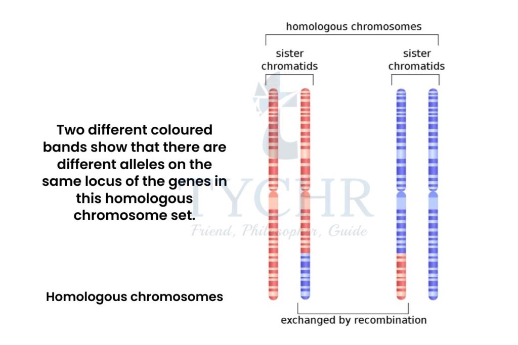
Figure 3.1 Homologous chromosomes
Ploidy of the cells
- This is the number of sets of chromosomes that exist in a cell.
- Types:
- Haploid (n); having only one set of chromosomes
- Diploid (2n); having two sets of chromosomes
- Triploid (3n); having three sets of chromosomes
- A human cell is a diploid cell (2n), with n = 23 i.e. 2n = 46 chromosomes, while their gamete cell is haploid (n).
- The sets of chromosomes varies in different species and so there ploidy changes.
Karyograms and Karyotypes Karyotype is the picture of the chromosome of an individual and karyogram is the standard representation of the chromosomes in the order, according to shapes and sizes. |
Sex determination
- The 23rd pair of chromosome is sex chromosome, as it determines the sex of the individual.
- There are two sex chromosomes namely; ‘Y’ chromosome and ‘X’ chromosome. Y- chromosome being much smaller than X-chromosome contains much lesser amount of genes.
- In human females, there are two X-chromosomes and the gamete produced by them will have only X-chromosome. While the males have one Y-chromosome and one X- chromosome with the chances of either of them getting into their gametes.
- Half of the sperms produced by male have Y-chromosome and the other half contains X-chromosome. So, when they fuse with the egg cell (female gamete), there is a 50% chance of either of the chromosome to get fused.
- Therefore there is a 50% chance to have a baby boy and 50% chance to have a baby girl.
- The other 22 pairs of chromosomes (other than sex chromosome) are called autosomes.
Meiosis
Meaning and process of meiosis
- This is a special type of cell division which reduces the chromosomal content of a cell into exactly half of the original after its division, also called reduction division.
- It is required for the formation of gametes. As gametes have half of the genetic material from the other autosomal cells.
- A diploid individual has one copy of chromosome from father and one copy from The gametes produced by the parent have to be half each to restore the full content in their offspring.
Process follows:
1.) This process produces 4 haploid cells (n) from a single parent cell (2n).
2.) Before meiosis, mitosis occurs and gives rise to a cell with 2n/4 chromosomes.
3.) Since there has to be total four cells, therefore the cell undergoes meiosis two times.
Meiosis I
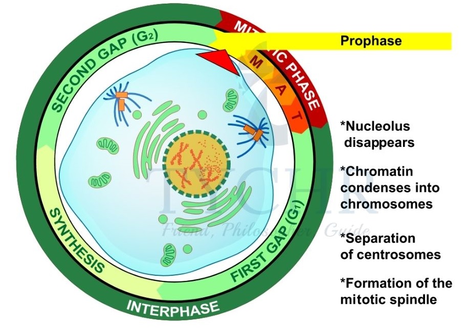
Prophase I
- DNA becomes compacted into visible chromosomal form.
- Homologous chromosomes attract each other inside the cell and pairs up.
- Formation of spindle fibers.
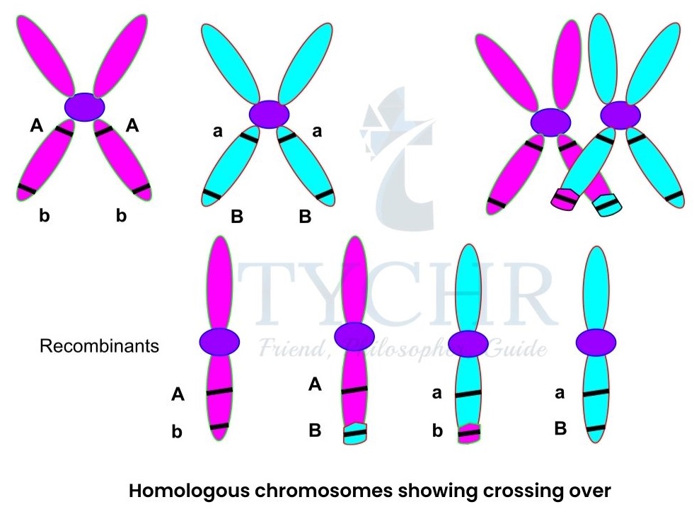
- Crossing over occurs, in which the non-sister chromatids of the homologous chromosomes exchange their genetic material to bring about the variation.
- Crossing over is an enzyme mediated process and the enzyme involved is recombinase. It allows DNA from a person’s maternal chromosomes to mix with DNA from the paternal chromosomes.
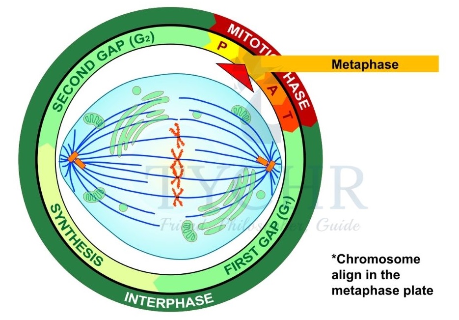
Metaphase I
- Homologous chromosomes line up at the equator of the cell by random orientation. This random orientation allows the combination to be different all the times the gametes produce i.e. responsible for variation.
- Nuclear membrane disintegrates.
Anaphase I
- Spindle fibres from the opposite poles, attach to chromosomes and pulls them towards the poles.
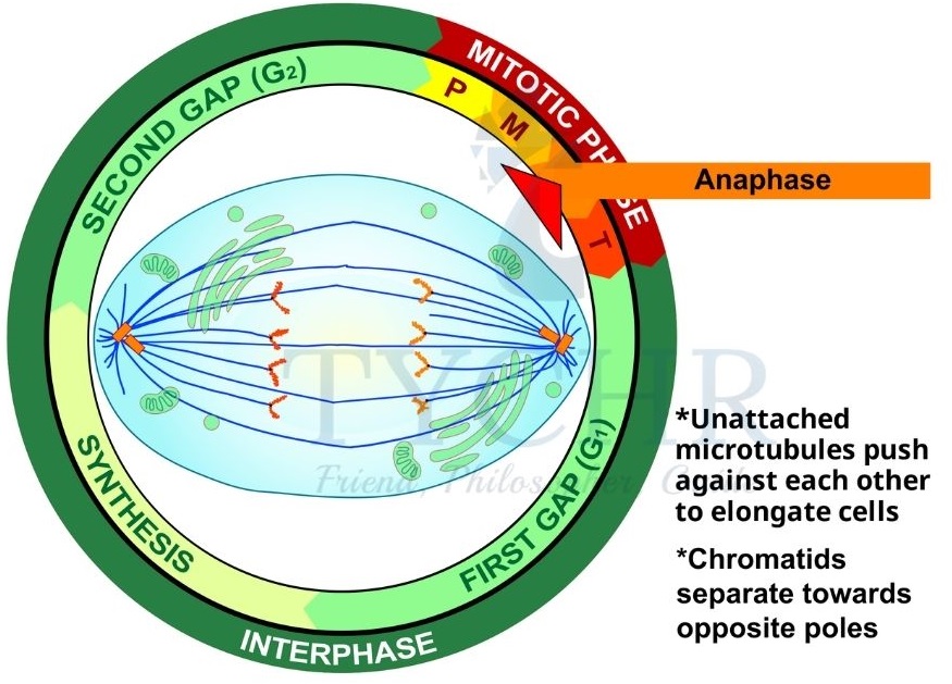
- Spindle fibres from the opposite poles, attach to chromosomes and pulls them towards the poles.
Telophase I
- Spindles and spindle fibers disintegrate.
- Nuclear membranes reform.
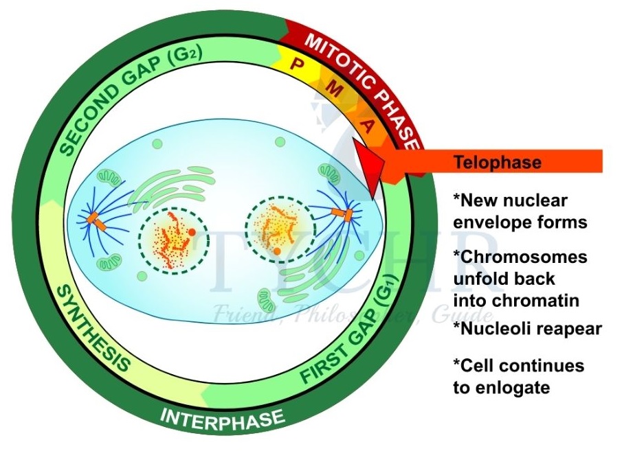
4.) Telophase is followed by cytokinesis and the cell splits into two.
5.) The two new cells produced are haploids with one chromosome of each pair.
6.) Each chromatid has its sister chromatids so no additional synthesising of DNA required and therefore the process continues.
2) Meiosis II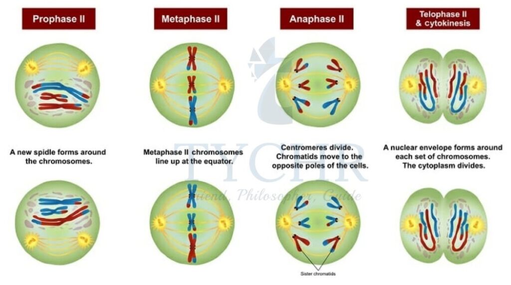
Prophase II
- DNA condenses again.
- New spindle fibers are formed.
Metaphase II
- Nuclear membrane disintegrates.
- Chromosomes line up on equator with random orientation again.
- Spindle fibers get attached to the chromosomes such that each spindle fiber from opposite poles attaches centromeres of each chromatid.
Anaphase II
- Spindle fibres contract and centromeres of each chromosome split, releasing each sister chromatid as an individual chromosome.
- The chromatids are pulled towards the opposite poles of the cell.
Telophase II
- Chromosomes uncoiled.
- Nuclear envelops are formed around each of the haploid four cells to undergo cytokinesis.
Chromosomal disorders
These are the disorders occur due to error in meiosis, which sometimes results in more or less chromosome number than the normal chromosome number.
Down’s syndrome is one of the disorders and caused by an additional copy of chromosome number 21 i.e. when the 21st chromosome fails to separate during Anaphase I, creating trisomy of the 21st chromosomes in the offspring.
Inheritance
Mechanism of inheritance
- Concept of inheritance was introduced by Gregor Mendel in 1865. He studied Pisum sativum (pea plants) for 10 years and brought out the conclusive pattern of inheritance.
- Pea plants have 7 contrasting characters including height, colour of pods, colour of flowers etc. Mendel crossed the pea plants by artificial pollination for a conclusion about all of these contrasting characters in every generation.
- Gametes receive one of the two alleles of a specific gene and this allele could be dominant or recessive with respect to the other allele.
- Dominant alleles: These alleles always express themselves in the phenotype over the other.
- Recessive alleles: These are the alleles which get masked by the dominant allele i.e. can’t express themselves in the presence of dominant allele.
Example: Let us assume two genes have two different types of alleles ‘AA’ (dominant) and ‘aa’ (recessive) which code for purple and white colour flowers respectively in a pea plant.Now, a pea plant with dominant ‘AA’ pair of alleles, it will show purple colour flowers and if it has recessive ‘aa’ pair of alleles then it will show white colour flowers
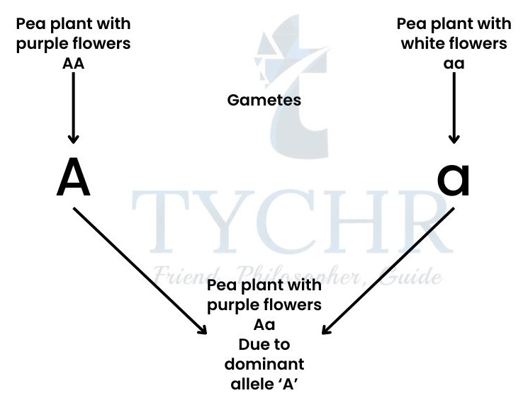
- This is called monohybrid cross because it includes only one contrasting trait i.e. colour of the flower and it concludes that, the recessive allele will only be expressed, when it is present in homozygous condition like in pea plant with white flower in the above case.
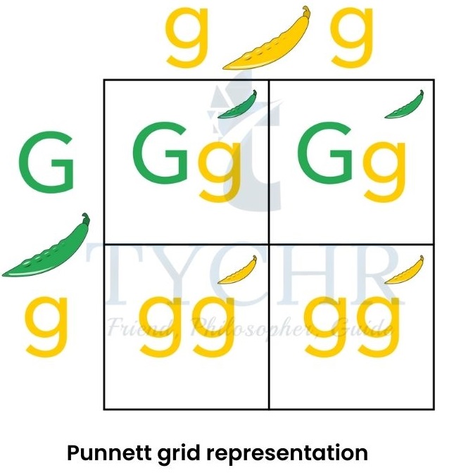
- To find out all the possible genotypes of the offspring that can be formed, we can use Punnett square or Punnet grid. Let us take two parents with heterozygous alleles ‘Aa’,
- This cross was first done by Mendel taking contrasting trait of height of the pea plant (tall or dwarf). ‘TT’ is the genotype of tall plants and ‘tt’ is the genotype of dwarf plants. He concluded, 75% of the offspring comes out to be tall and 25% comes out to be dwarf. This ratio 3:1 is the result of dominant and recessive allelic pairs. 50% of tall plants have heterozygous genotype (Tt) and 25% of tall plants and dwarf plants each, found to be of homozygous dominant and recessive genotypes, respectively.
Test cross
- This type of cross is done to determine the genotype of an unknown plant.
- The genotype of the unknown plant is crossed with homozygous recessive genotypic plant and the conclusions are carried out.
- If we cross Tt (heterozygous genotype) with tt (homozygous recessive), then we get Tt and tt in the ratio 1:1. But if we cross homozygous dominant TT with homozygous recessive tt then we get all the plants with heterozygous dominant genotype (Tt).
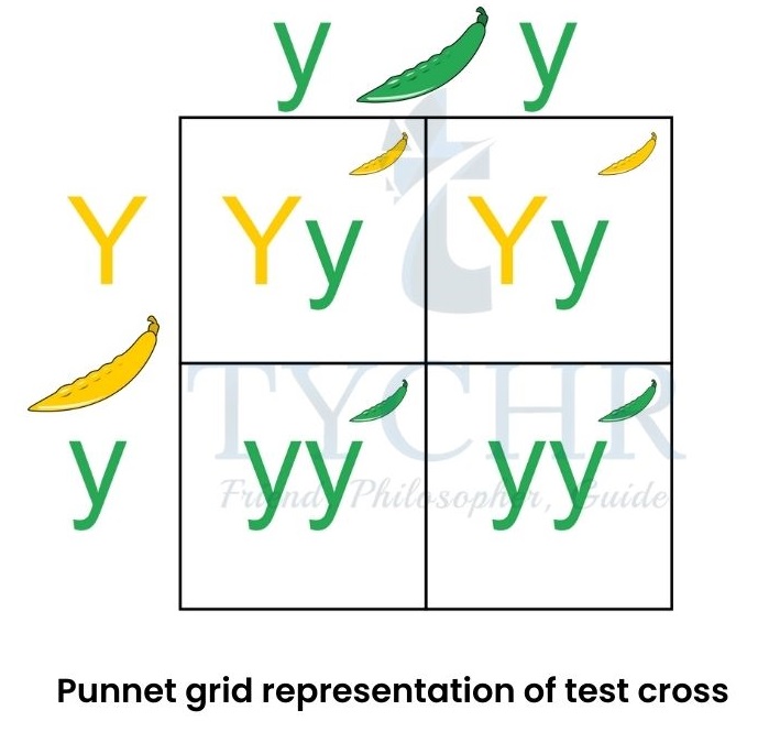
Figure 3.4 Punnet grid representation of test cross
Multiple alleles
There can be more than two alleles for the same gene like in the ABO blood grouping system of humans.
- ABO blood grouping system of the humans is determined by three types of alleles;
IA | codes for type A antigen proteins and gives type A blood |
IB | codes for type B antigen proteins and given type B blood |
i | this is a recessive allele, codes for none of the proteins and gives type O blood |
- Total six genotypes are formed while crossing all of these alleles together:
| IAIA | Type A blood |
| IAi | Type A blood |
| IBIB | Type B blood |
| IBi | Type B blood |
| IAIB | Type AB blood |
| ii | Type O blood |
- Type AB blood group shows codominance.
- If parents have IAi and IBi genotypes respectively, then they can have 4 children with different blood groups
Genetic diseases
There are two kinds of genetic diseases in humans.
- Autosomal genetic disease
- The disease caused by alteration in genetic material of autosomal cells rather than sex cells is called autosomal genetic disease.
- They are caused by recessive types of alleles which are found in the first 22 pairs of chromosomes but not in any of the X or Y sex chromosomes.
- Examples are: albinism, phenylketonuria (PKU), cystic fibrosis, sickle cell disease, Tay Sachs disease, and thalassemia.
- Sex-linked genetic disorder
- The disease caused by alteration in an allele found on either of the sex chromosomes X or Y is called as sex-linked genetic disease.
- Y chromosome is much smaller than the X chromosome and therefore some locus are present on the X-chromosome has nothing to pair up with.
- Examples include: colour blindness and haemophilia. People can’t differentiate between green and red colour when have colour blindness and blood clotting does not occur while suffering from haemophilia. Alleles for both these diseases are found on X-chromosome.
- Xb is the allele for colour blindness while XB is for normal vision.
- Xh is the allele for haemophilia while XH is for normal.
- No allele is present on the Y-chromosome.
Possible genotypes for being affected or not affected XBXB or XHXH
non-affected female
XBXb or XHXh
non-affected female but a carrier
XbXb or XhXh
affected female
XBY or XHY
non-affected male
XbY or XhY
affected male
- Carrier female is the one who can pass the affected allele to the next generation. Their affected allele is masked by the dominant normal allele.
Biotechnology and genetic modifications
Gel electrophoresis
- This technique is used to separate DNA fragments (chopped out of long DNA strands by the action of enzymes), according to their sizes.
- An apparatus set for this technique is constitute of gel (generally agarose gel) which provides sieve and two ends with positive and negative charged plates.
- DNA fragments are kept in the gel and when an electric current is passed the negatively charged DNA gets to move towards the positive end of the apparatus by passing through the sieve gel.
- Accordingly, the DNA fragments gets separated by moving towards the positive end with the lighter one travelling larger distance and heavier segment of DNA travelling less.
- A DNA probe, which is a small segment of complementary DNA, is added during the process and it gets attached to the gene of interest.
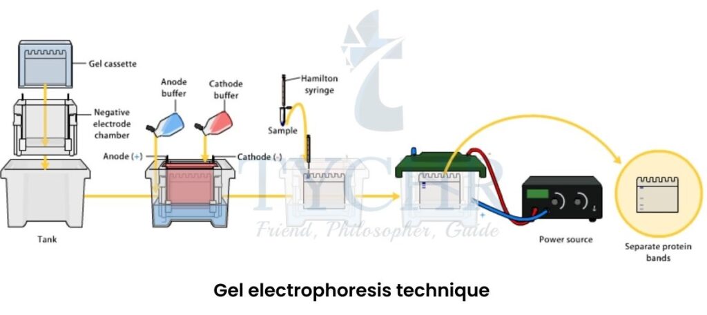
Figure 3.5 Gel electrophoresis technique
Polymerase chain reaction (PCR)
- This technique is used to amplify the gene of interest obtained from gel electrophoresis, using thermocycler.
- A very small amount of DNA segments are inserted into the thermocycler and a huge volume of it can be obtained.
- Large quantities of DNA segments are required to analyse it efficiently with many trials.
- A special enzyme taq-polymerase is used, it can withhold extreme temperatures.
DNA profiling
- This is the process for matching an unknown sample of DNA with a known sample to see if they correspond. It is also called DNA fingerprinting.
- After gel electrophoresis, if any two DNA fragments are found to be identical in bands of autoradiograph, then that can be regarded as a match.
- Applications of DNA profiling:
- To identify someone’s biological father by matching the DNA of the child with the
- Can be helpful in crime scenes such as
- Study the ecosystem by taking DNA samples from different species around and finding the ecological similarities between
- Analysation of DNA profiling is done by examining the autoradiograph made by gel
Figure 3.6 Autoradiograph showing banded lines for DNA profiling
Genetic modifications
Gene transfer
- The technique of transferring a gene of interest from one organism (donor organism) to other organism (host organism) to modify them in search of availing more benefits, also called as recombinant DNA technology.
- Genetic modifications are carried out by this technique.
- This technique is possible due to the fact that all the organisms have the same genetic makeup.
- Examples include: Bt corn and Bt cotton. A soil bacterium named Bacillus thuringiensis produces a certain type of protein that causes formation of pores in the alkaline gut of bugs and ultimately leads to the death of bugs.. This protein encoding gene was extracted from bacterium and transferred into the DNA of the crops like corn and cotton, making them resistant to bugs.
Clones
- These are the exact copy of the individuals already exist, made by using genetic techniques which are;
- Cutting of the DNA using endonucleases enzymes. These are restriction enzymes which cut the DNA at specific sequences only. This results in a DNA segment or gene of interest to be separated out of the whole chain.
- This gene is removed out from the donor organism and two ends of the DNA which hangs (sticky ends), gets pasted by the enzyme named DNA ligase.
- Now the gene of interest is glued to the plasmid of the host cell to make its copies. Host cell is mostly bacterium E.coli due to its short lifetime (it doubles every 20 minutes), so that we can get lots of copies in short period of time.
- Host bacterium is kept in bioreactors to provide them with the appropriate conditions for copying (cloning of gene).
- This technique is used to produce artificial insulin in the laboratories.
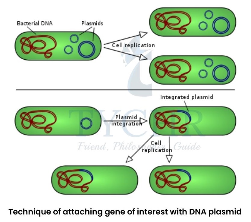
Figure 2.7 Technique of attaching gene of interest with DNA plasmid
Genetically modified organism (GMO)
GMOs are called transgenic plants or transgenic animals. The organism is modified such that the undesired traits are masked, whether by removing the undesirable gene and adding the new or by adding the new gene only.
Transgenic plants
- First GM plant which was commercially available was Flavr savr tomato. This was genetically modified to delay ripening and rotting and to stay fresh for longer.
- The rice plant was genetically modified for production of beta carotene in rice grains, to eradicate the deficiency of vitamin-A.
Transgenic animals
- The animals are genetically modified to produce a substance that can be used in medical treatments.
- A sheep was genetically modified to produce milk with the protein called factor IX, which is helpful for the treatment of haemophilic patients.
- First transgenic cow Rosie, produced milk with alpha-lactalbumin which is more balanced product for babies.
Dolly – the sheep A sheep named “Dolly” was introduced in 1996 by the Roslin Institute in Scotland. She was the animal clone made out of somatic cell nuclear transfer technique. The technique follows as: |