animal physiology Notes
Animal physiology
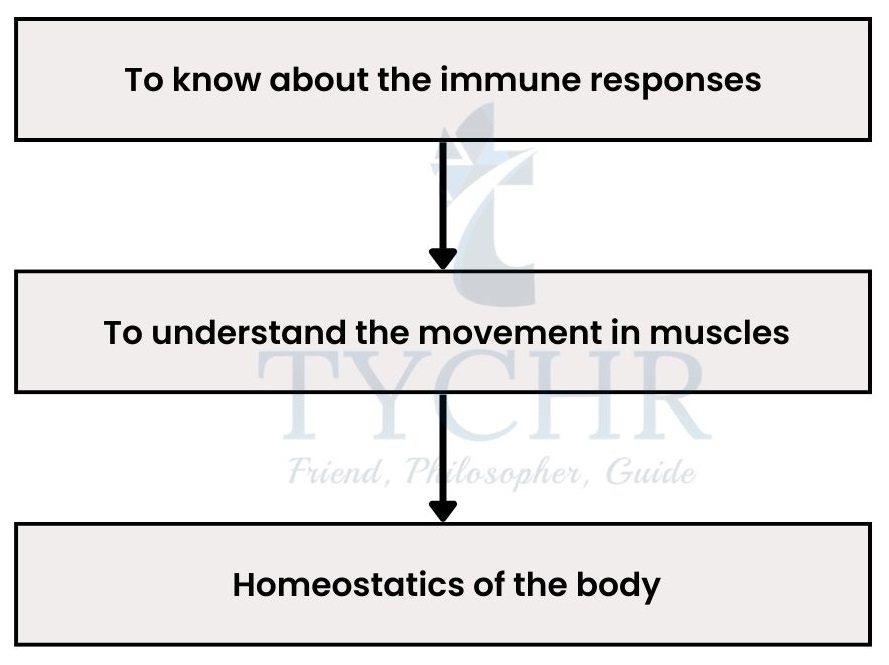
Approaching the topics
- Understanding the concepts
- Application of these concepts
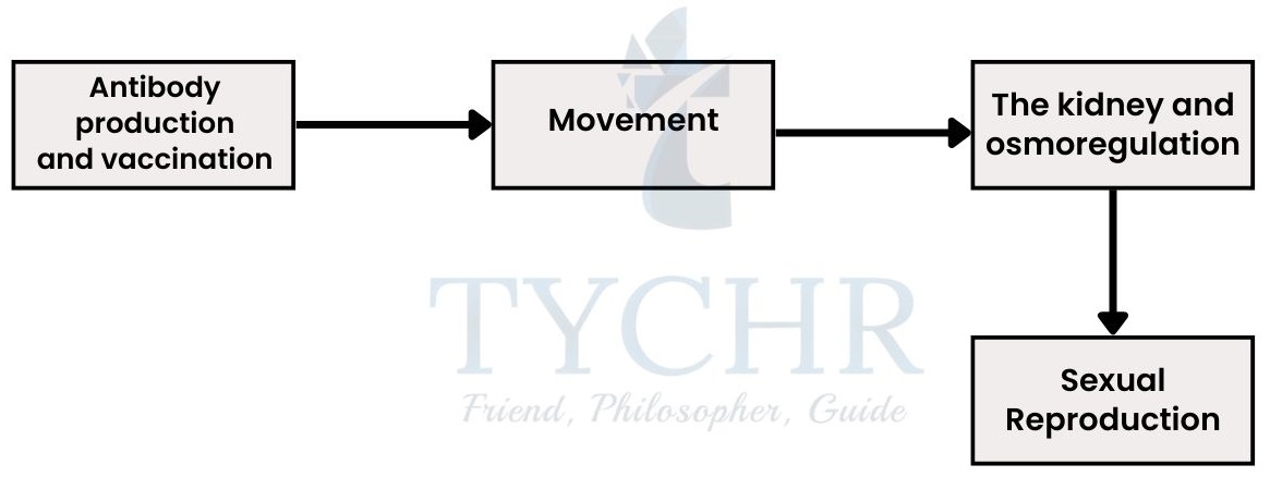
Antibody production and vaccination
Immune response
- Immune system recognizes any foreign cell through antigens.
- On the cell’s surface, antigens are proteins and polysaccharides.
- Invading viruses, bacteria, fungi and even transplanted organs or blood that is transfused all have different plasma proteins or antigens.
- An immune response is generated against the invading cells (recognised as not-self by leucocytes).
- Immune response follows:
- Macrophages: These are massive leukocyte cells, which engulfs the pathogen by the process called phagocytosis. And present antigen of the pathogen to their cell membrane (antigen presentation).
- T lymphocytes recognise the antigen presentation and become activated and now the immune response is antigen-specific.
- T lymphocytes bind to B lymphocytes and simulate them to produce specific antibodies which further attach to the antigen by their binding sites and deactivate them.
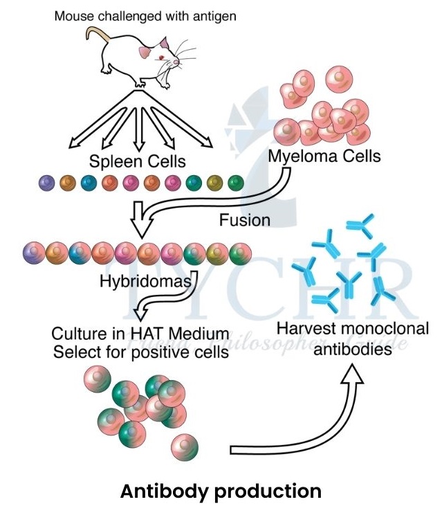
Figure 11.1 Antibody production - B cells are less in number while initiating the response but they clone themselves (undergo mitosis) to produce large amounts of antibodies. They have a rough endoplasmic reticulum (rER) which produces and transports antibodies. Some of these plasma cells become memory cells (long lived) which eventually produce secondary response.
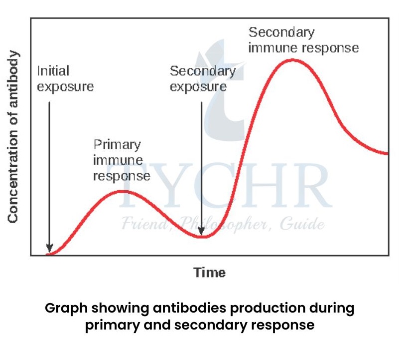
- Memory cells stream in blood vessels but remain inactive until the pathogen it is specific to, attacks again. They become active if the same type of pathogen invades again and starts producing the antibodies before the symptoms are evident.
Blood transfusion and how it is unique?
- We use the ABO blood grouping system, and each blood type contains distinct antigens;
Antigens are present in Type A, Type B, and Type AB blood cells, respectively, while there is no antigen present in Type O blood cells. - One more protein is present in the blood called Rh protein, which differentiates it into two categories; Rh positive and Rh negative based on the presence and absence of it. And according to presence and absence of Rh factor, the blood types are notated as X+/X–, ‘X’ may be A, B, AB, O.
- If the donor and recipient both have different blood types, then the blood transfused into the recipient’s body will cause an immune response from the recipient’s immune system, due to different types of antigen (not-self) on the donor’s blood cells.
Vaccine
- A vaccine contains attenuated or killed pathogens or their derivatives to generate the primary response in human’s body against the pathogen, so that if it attacks in actual secondary immune response could be stimulated for rapid cleaning of pathogens.
Monoclonal antibodies ̶ Production and use
- Monoclonal antibodies are specific antibodies for only one type of antigens i.e. they are exactly the same.
- Primary response in the organism is polyclonal response because of the presence of different types of antigens on the pathogens.
- Production of monoclonal antibodies:
- An antigen (of interest, against which we want to produce antibodies) is injected into the mammal or a mouse.
- Primary response is generated inside the animal i.e. plasma B cells are produced. Their spleen is taken out and the B cells are taken out.
- B cells are fused with cancer cells called myeloma cells so that the leukocytes remain alive for a long time. Hybridoma cells (hybrids of antibody-producing cells and long-lived myeloma cells) are formed when B cells and myeloma cells fuse.
- Pregnancy test results are a colour indicator, why?
- Embryo inside the womb secretes hCG (human chorionic gonadotropin) hormone which is added into the bloodstream. Hybridoma cells can be formed and anti-hCG antibodies bonded with an enzyme will produce colour when encounters with hCG molecules.
Wonder! What if, the immune system itself starts to cause harm to the body? The function of immune system is to generate response against the foreign invader in the body through any external source. It works on the basis of self or not-self component at major, not on harmful or not harmful. The allergens like pollen, egg white, peanuts etc. are harmless but are identified as not-self and therefore an immune response is generated against them. Particularly IgE antibodies are released which bind to mast cells and the allergens as well, it triggers mast cells to release histamines. The histamines are the chemical which causes allergy symptoms like sneezing, congestion, itching, red skin etc. i.e. histamines are the cause of allergy. |
Movement
Bones and skeleton
- Bones together make up a structure called skeleton, it can be made up of bones or chitin. On the basis of its composition skeleton can be of two types:
- Endoskeleton: It is made up of bones and provides structural support in vertebrate animals.
- Exoskeleton: It is made up of chitin and provides structural support in insects. Skeleton works with muscles and therefore the endoskeleton has muscle attachment points on its outside and the exoskeleton has muscle attachment points on the inside.
- Some bones act as levers to facilitate movements of some bony parts. It works in the mechanism and includes a fulcrum between the effort force and resultant force. The levers have three classes depending upon the positions of the three components;
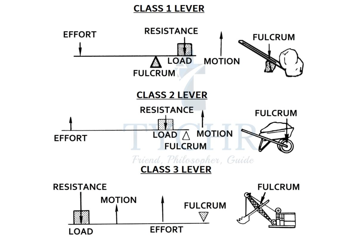
- Spine is considered a first-class lever by perfectly lifting the chin through the action and reactions of effort and reaction force.
Working of muscles
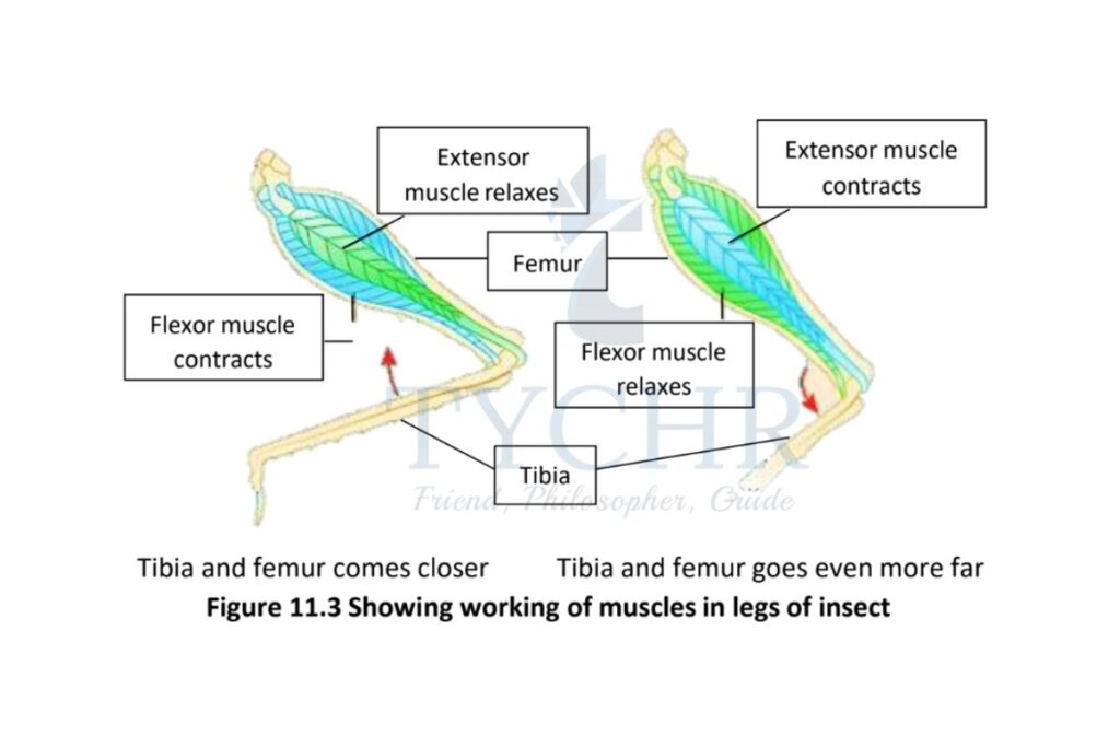
- Muscles work antagonistically in pairs to contract and relax at the same time, causing movement of the bone.
- In a pair of muscles; one is connected to the part of the immovable bone and the other is connected to the movable part of the bone. One muscle contracts and the other relaxes (antagonistic) to cause a movement.
- Example of muscle working in the insect legs:
- Movement of muscles causes movements of bones in the skeleton of mammals through the joints. These bone-to-bone joints are:
- Synovial joints: These are the joints with a lubricant called synovial fluid.
Ex – Human elbow - Ball and socket joint: It includes the joint of the humerus and shoulder which provides movement in various directions. It is also the joint between femur and pelvic girdle in the legs.
- Hinge joint: It is one of the synovial joints which provides restricted movement along the axis. Ex- knee joint and elbow joint.
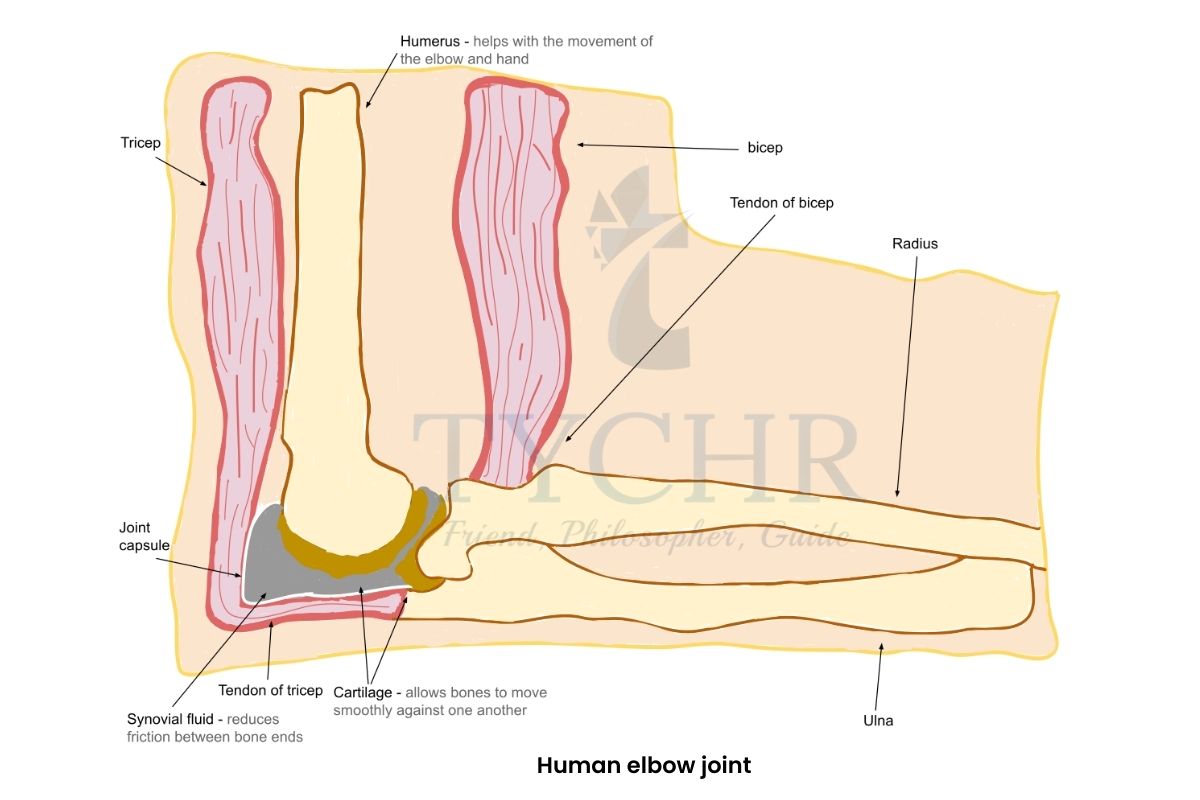
Figure 11.4 Human elbow joint
- Synovial joints: These are the joints with a lubricant called synovial fluid.
Structure and action of skeletal muscles (striated muscle)
- Muscle fibre
- Muscle is composed of thousands of cells called muscle fibre. They are multinucleated
and have a specialised endoplasmic reticulum.
- Muscle is composed of thousands of cells called muscle fibre. They are multinucleated
- Plasma membrane of these cells is called sarcolemma, it has transverse tubules or T tubules penetrating into the cell.
- The cytoplasm of these muscle cells is called sarcoplasm which contains many organelles and a molecule called myoglobin (just like haemoglobin), it stores oxygen and supplies it when not enough oxygen is provided by haemoglobin like when we are exercise.
- There are many parallel elongated structures called myofibrils inside the muscle fibre which is packed with the mitochondria to provide ATP at the time of muscle contraction.
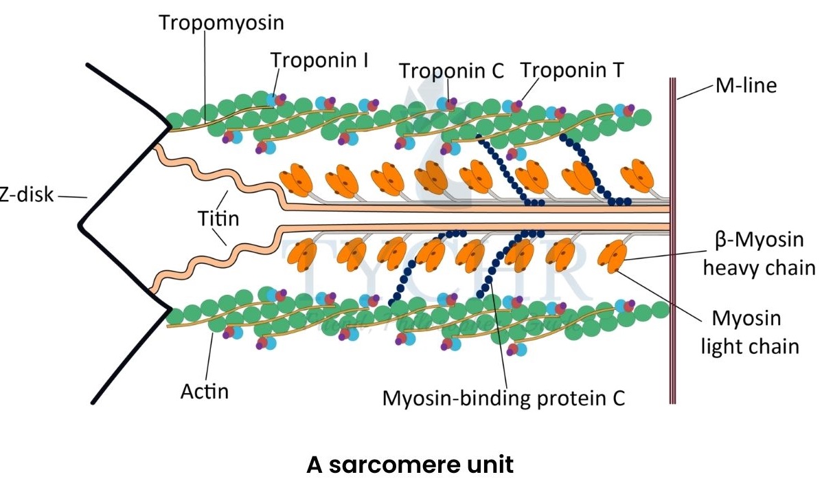
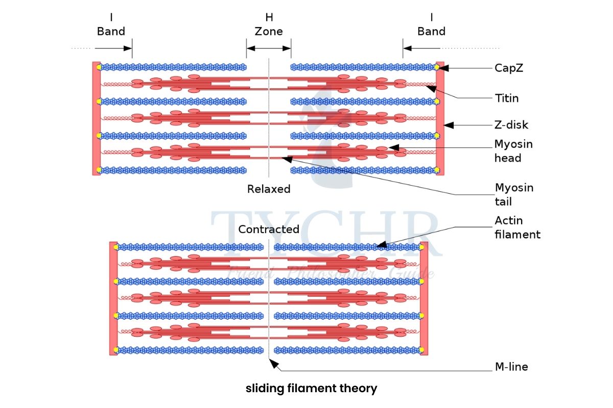
Figure 11.5 A sarcomere unit - Myofibril is made up of sarcomere units which give it dark and light bands. Each sarcomere is composed of proteins actin and myosin.
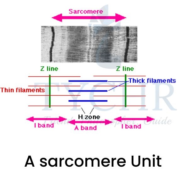
- Myosin gives dark shade and actin gives light shade. Actin in each sarcomere slides
towards the H zone and brings about the contraction of one sarcomere.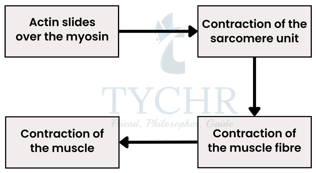
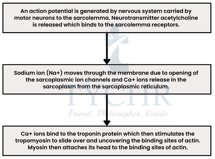
Sliding filament theory ̶ Mechanism of muscle contraction
The Kidney and osmoregulation
Nitrogenous wastes
- There are three types of nitrogenous wastes; ammonia, urea and uric acid.
- Ammonia: It is very toxic in blood and tissues therefore must be diluted with much water and removed out of the body. It is a waste product of fishes.
- Urea: It is also toxic in blood and tissues and therefore diluted with water and removed out of the body. It is found as a waste product of mammals.
- Uric acid: It is found in birds as a waste material. It is insoluble in aqueous solution such as blood, cytoplasm etc. It requires less or no water for dilution and removed out from the
Organisms have developed there physiological functions such like that to produce different types of waste material but composition of nitrogen. Like fishes can access a lot of water so they can go easy with ammonia, mammals can have sufficient amount of water accessible to them to pass out urea and birds fly in the sky and have beaks with not much affinity to get in water, therefore uric acid.
- Energy requirement to produce uric acid is much more than used for the production of urea and then to produce ammonia.
Nitrogenous wastes in insects and its excretion
- Insects have a specialised structure called Malpighian tubules. These tubules are close from one end and open from the other, the blood travels inside the tube and undergoes selective reabsorption.
- The nitrogenous wastes (uric acid), excess water and ions remain in the tubules and useful nutrients and water gets absorbed.
- The Malpighian tubules end in the gut and waste ultimately gets out of the body into faeces.
The kidney ̶ its anatomy
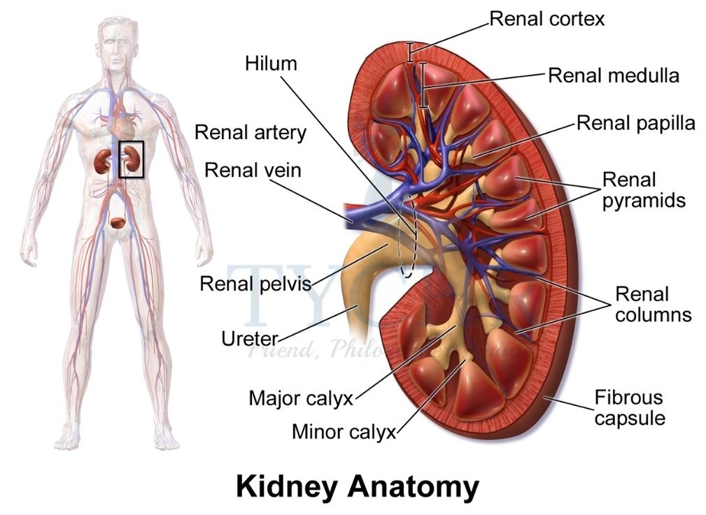
- Kidneys filter blood and separate out the waste products. It produces urine which is a combination liquid of water and waste products (from the blood).
- After formation, urine collects in the renal pelvis of the kidney which drains it into the ureter and further urine gets (stored) into the urinary bladder until it gets full.
Where in kidney does filtration occurs? - Nephrons are the constituents of the kidney. These are the small filtering units which are present in more than a million (in number) in each of the kidneys.
Nephron structure and mechanism of function
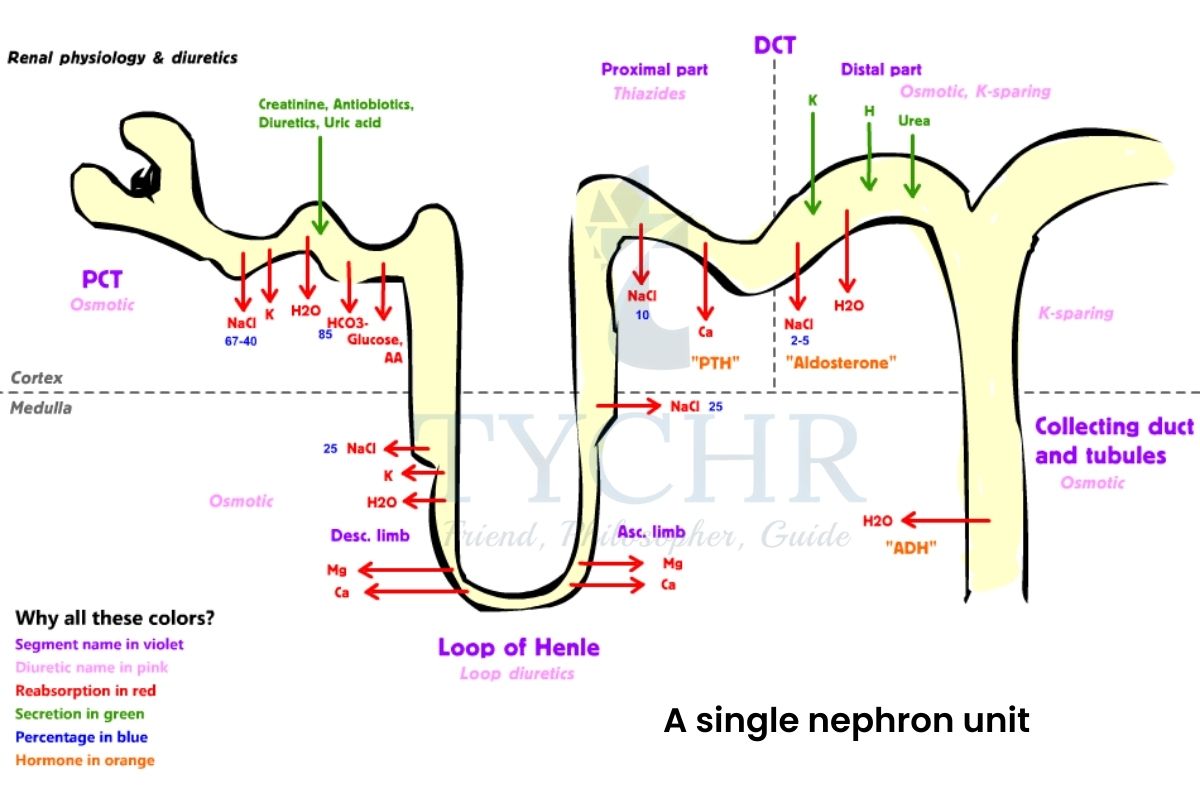
- Renal artery brings unfiltered blood to the kidney and renal vein take away filtered blood away from the kidney.
- Parts of nephron; glomerulus, Bowman’s capsule (surrounding the glomerulus), proximal convolutes tubule (PCT), loop of Henle, distal convoluted tubule (DCT) and a peritubular capillary bed surrounding these tubules.
- Each nephron shares both parts of kidney (cortex and medulla).
- Blood from the afferent arteriole enters the glomerulus (capillary bed with fenestrations) which tends to increases the pressure of the blood once it enters it.
- Blood circulates in the glomerulus fenestrated capillary bed and exits from the efferent arteriole. The filtrate is passed on to the tubules part of the nephron.
- The filtrate obtained after passing through glomerulus is ultra-filtered, allowing glucose molecules but not protein, to pass through.
- First of all, the filtrate enters the proximal convoluted tubule (PCT) from where the reabsorption of valid substances begins.
Reabsorption
- Filtrate flows into the lumen of single-celled thick PCT which is lined by microvilli for better reabsorption.
- Salt ions (by active transport), required water (by osmosis), glucose (by facilitated diffusion) moved out of the PCT and carried away by surrounding peritubular capillary bed.
Osmoregulation
- The response mechanism of the body to maintain the water balance is called osmoregulation.
- While water is still there in the filtrate to make it travel through the rest of the nephron, it enters the loop of Henle.
- Loop of Henle has two portions; descending portion (it is permeable to water but impermeable to ions) and ascending portion (it is impermeable to water but permeable to ions).
- Therefore some water gets out in the descending portion, due to hypertonic region medulla, and some salt ions move out in the ascending portion.
- The filtrate still remains hypotonic when it reaches DCT, and therefore a hormone ADH (anti-diuretic hormone) comes into play to reabsorb some more water in the collecting tubule.
- ADH is secreted from the posterior pituitary and makes collecting duct permeable to water so that excess water moves out due to osmosis.
Osmoregulators and osmoconformers
- The animals which are having different solute concentrations compares with that of environment are called osmoregulators. They have built-in mechanisms to regulate water balance by the expenditure of energy.
- The animals which have same solute concentrations as that of surrounding environment are called osmoconformers. Since they are already maintained, they do not exhibit any mechanism of regulations. Water from them moves in and out freely due to the same concentrations inside and outside.
Sexual reproduction
Spermatogenesis
- The process of formation of spermatozoa (sperm cells) is called spermatogenesis. The process occurs in the small tubes called seminiferous tubules inside the testes.
- Germinal epithelial cells (spermatogonia) lie near the outer wall of the seminiferous tubule which undergoes meiotic and mitotic division to produce spermatozoa.
- Steps follows:
- Diploid spermatogonia (having 46 chromosomes) undergoes meiotic I division and results in two haploid cells (23 chromosomes each).
- Each haploid cell then undergoes 2nd meiotic division and produces four haploid cells which mature into spermatozoa.
- Each spermatozoon remains in the seminiferous tubules until it matures and during this time they get nutrients by embedded their heads in Sertoli cells.
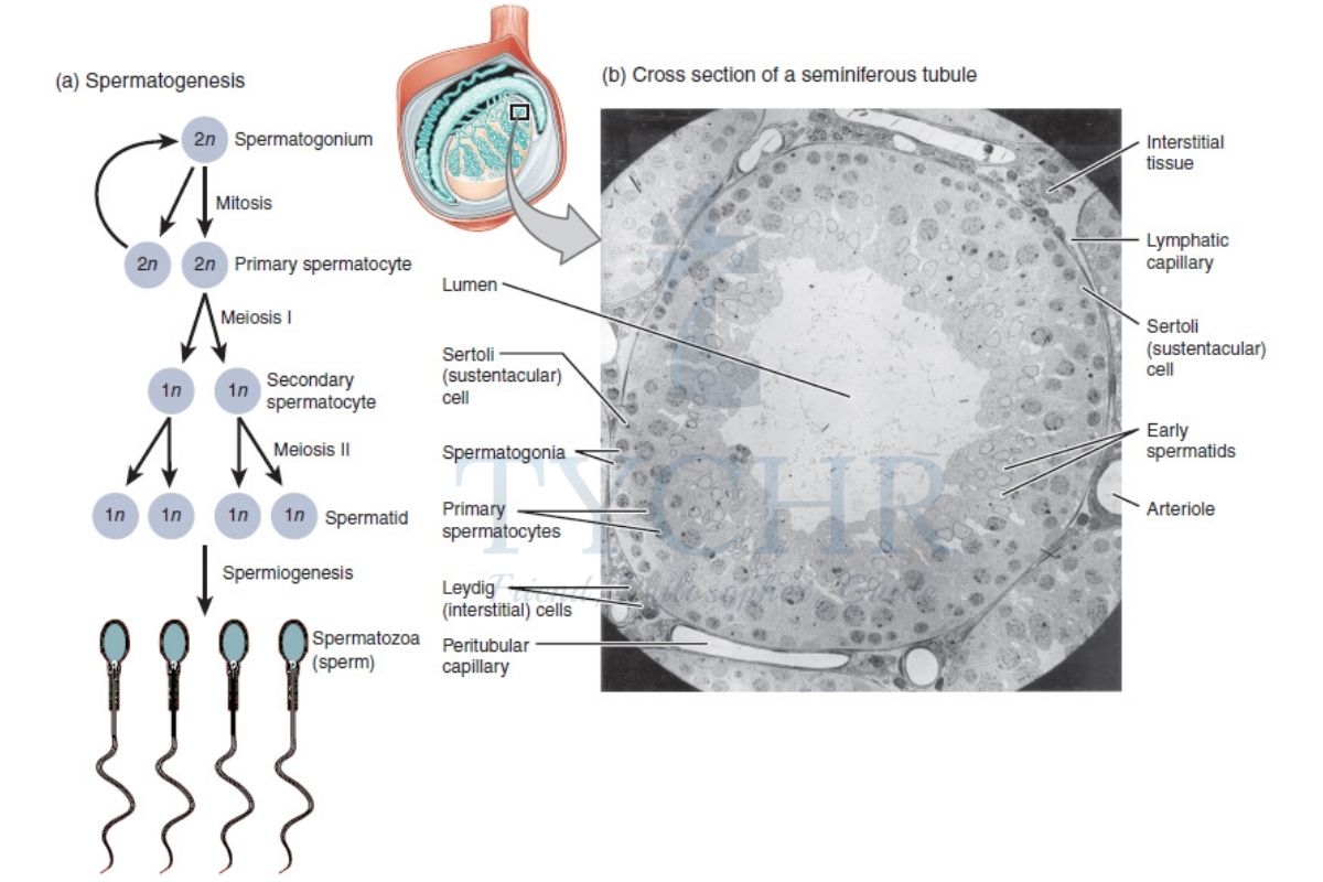
Figure 11.9 Process of spermatogenesis - Once fully mature the sperm cells move into the epididymis.
Oogenesis
- The process of formation of ovum is called oogenesis.
- The process starts when the girl child is still in the womb. Oogonia present in the ovaries starts to grow into a mature form called primary oocytes. Primary oocytes undergo prophase I stage of meiosis and get arrested at that stage.
- Follicle cells inside the ovary also undergo mitotic division which further surrounds the primary oocyte and the overall structure is called primary follicle.
- After attaining puberty, cyclic changes start and during each menstrual cycle primary follicle completes meiosis I and results in one large secondary oocyte and polar body.
- Secondary oocyte is of much larger size than the polar body (it is just containing half of the chromosome).
- The follicle outer ring starts to divide into two and fluid gets filled between, with the inner ring surrounding the oocyte. This form of follicle is called Graafian follicle.
- The secondary oocyte (ovum) is released from the ovary and it will complete its second meiotic division only if it gets fertilised.
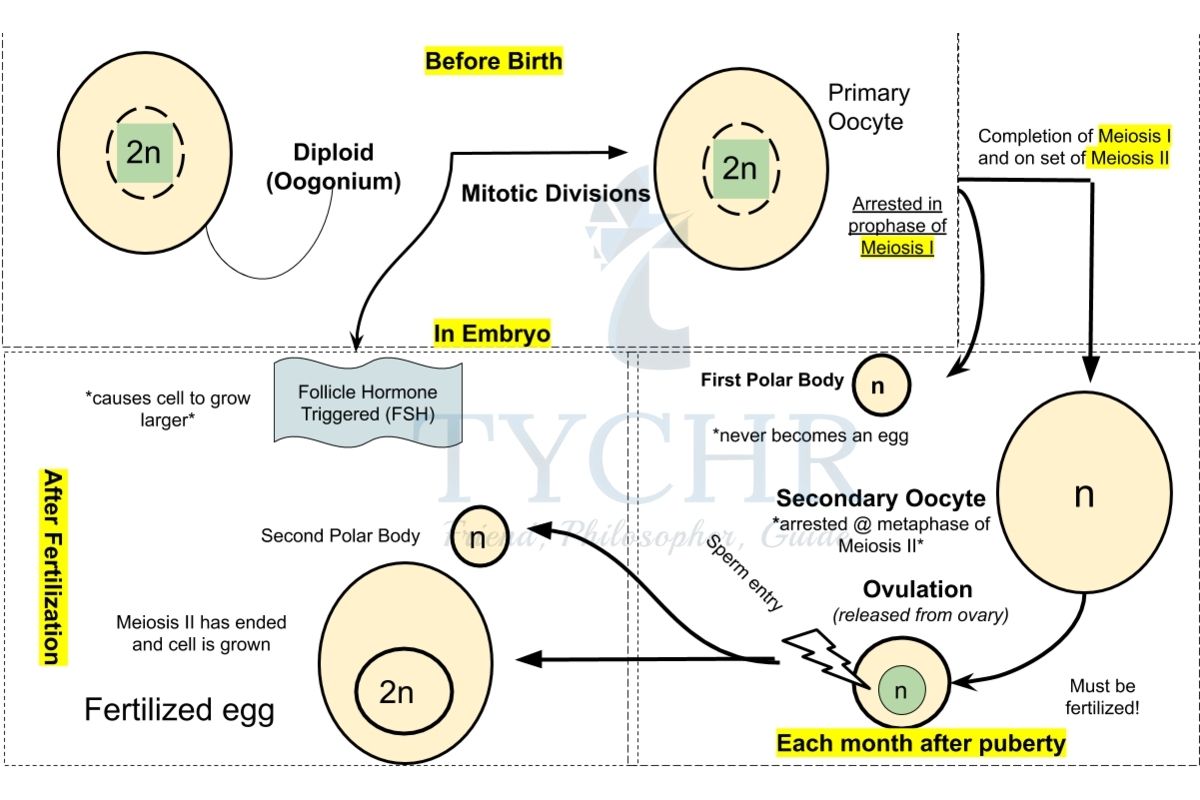
Figure 11.10 The process of oogenesis
Fertilization
- The fusion of the male and female gametes results in fertilisation. It could be external or internal.
- The process of external fertilisation involves releasing the male and female gametes outside of the female body through the use of a medium, typically water, where they fuse. This method of fertilisation is utilised by some amphibians and fishes.
- In internal fertilisation, the male gamete is released inside the body of the female and the male and female gametes fuse inside the female body. It occurs in mammals and reptiles etc.
Human fertilization
- Millions of sperms are ejaculated at the time of copulation, which travel into the fallopian tube where the egg is present to fertilise it.
- The egg is surrounded by follicle cell layer and zona pellucida and it takes many of the sperms to get through them.
- Only one sperm out of millions gets inside the ovum to fertilise it and this is made sure by the cortical reaction that occurs just after a sperm cell enters.
- The cortical reaction releases an enzyme which hardens the zona pellucida and makes it impermeable to the rest of the sperm cells.
Implantation
- After fertilization, mitotic division starts which increase the count of zygotic cell up to 100 within a week. It is now called blastocyst with a surrounding layer called trophoblast.
- At this stage it reached uterus from the fallopian tube, for implantation into Trophoblast releases some enzymes to embed into the endometrium and it also helps to form placenta.
- Till it gets embedded into the endometrium, the yolk nutrients of the egg gets used up but fortunately the placental connection between the mother and the embryo is made.
What does placental connection do?
- Placenta is formed from mother and embryonal tissue i.e. it is the only connection between the two.
- The exchange of fresh nutritive blood and deoxygenated blood with waste products occurs through the blood vessels in the umbilical cord of the placenta.
- There are total three blood vessels work to keep the baby healthy:
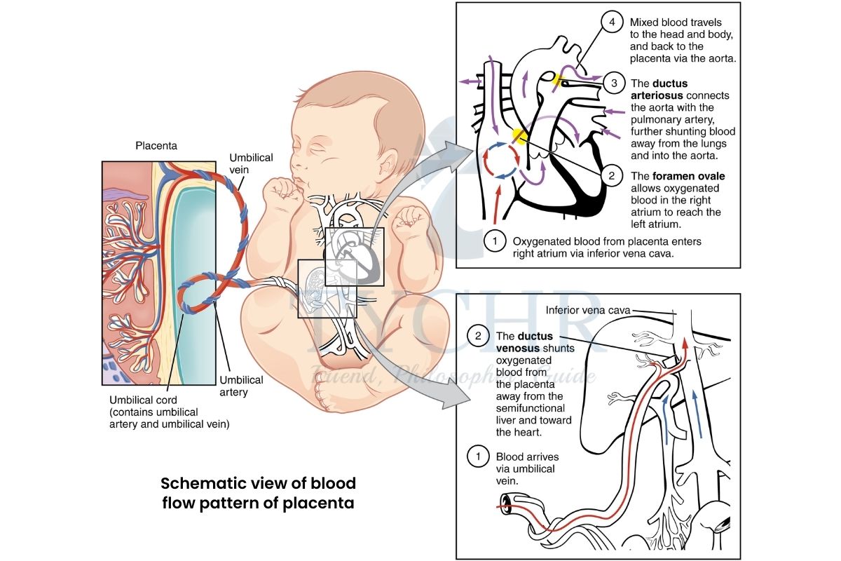
Figure 11.11 Schematic view of blood flow pattern of placenta
Hormonal balance
- Embryo secretes hCG hormone which enters the mother’s bloodstream and leads to further releases of progesterone to maintain the endometrium corpus luteum.
- But eventually, when corpus luteum degenerates, the placenta takes over the job and releases progesterone. It also releases oestrogen.
Parturition
- A small amount of oxytocin hormone is released by pituitary which causes uterine contractions.
- The contraction in turn signals to release more oxytocin i.e. contraction and production of oxytocin are leading each other.
- Uterine contractions over and over again, cause the baby to come out, which is called parturition.
Zoonosis Zoonosis is a pathogenic disease which transforms from any vertebrate animal to humans, like bird flu, Lyme disease, West Nile virus, Bubonic plague, Nipah virus, Covid-19 etc. These zoonotic diseases are becoming more familiar due to more and more contact of humans with wildlife or may be by disrupting wildlife. The transfer of zoonotic disease may be through direct contact like with air, bites or saliva and by eating them or by some vectors. Contaminated food may also be the cause of transfer. Human behaviour is also one of the main cause. Exploitation activities, deforestation etc. may increase the contact of animals with the humans. |

