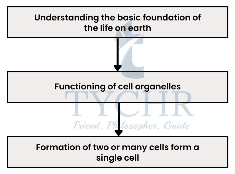
The cell is the fundamental unit of life. This study note explores the intricate world of cells, covering key concepts within the IBDP Biology syllabus, including cell structure and function, cell membranes, transport mechanisms, cell division, and cellular respiration. A thorough understanding of cell biology is crucial for success in IB Biology and for comprehending all biological processes.
Cells
- Understanding the topics

- Application of these concepts


Basic terms to ponder:-
- Microscope: An instrument used in laboratories for small substances that cannot be seen by the naked eye.
- Micrographs: A digital image or a photograph taken by a microscope.
- Phosphorylation: Addition or attachment of phosphoryl group to an organic compound or protein complex.
- Meristematic tissue: These are present at the tips/apex of roots or stems. Starting from a single apical cell, it becomes capable of forming the entire plant.
Cell
Cell theory
Cell is the smallest collective functional unit a living organism consists of. When a number of cells collectively perform a specific function, then it is called a tissue or an organ.
Scientists who were involved in discovery and recognition of the cell are:
- Robert Hooke in 1665, discovered the cell after observing the cork of a tree by his own microscope.
- Later, Antonie van Leeuwenhoek observed the first living cell.
- In 1838, Mathhais Schleiden stated that plants are made of cells.
- In 1839, Theodor Schwann made it clear that animals are made up of separate cells.
The contributions of these scientists made the solid foundation of the cell theory which has three principles:
- All the organisms are composed of one or more cells.
- Cells are the smallest units of life.
- All cells come from pre-existing cells.
Organisms exist either in a unicellular or multicellular form and perform certain necessary functions of life which are; metabolism, reproduction, homeostasis, excretion, growth, response to stimuli and nutrition.
Cell: Size and Structure
Every cell has its own smaller subunits which can be observed by using microscopes.
- Light microscope:
This microscope passes light through the specimen (either living or dead) to form an image. It is cheaper and can magnify up to 2000× from a normal vision. - Electron microscope:
This microscope passes electrons through dead specimen to form and image. It is expensive, complex and can magnify up to 500000× from a normal vision.
The order of size in the micro-world goes like:
organelles (of a human cell) > bacteria > viruses > membranes > molecules.
To find the magnification done by microscopes, scale bars are placed next to micrographs and the magnification is given by the formula:
magnification = size of image /size of specimen
- Surface area to volume relativity ratio limits the size of the cell;
- The greater the surface area of the cell, the higher movement of the materials in/out of the cell. The greater the volume of the cell, the greater the rate of heat and waste production, and the rate of resource consumption of the cell because chemical reactions occur inside the cell only.
- Large cells by volume have relatively less surface area per unit volume because volume increases by the factor of cubing the radius and surface area increases by the factor of squaring the radius.
- Some cells with folding can have larger surface area with respect to volume.
Differentiation of cells
- Single cell undergoes rapid reproduction to form a multicellular organism and further differentiation to produce all the required cell types.
- Specific genes are involved in the production of certain types of cells, with each cell having the complete genetic information of the organism.
Nerve and muscle cells, for example, can lose the ability to reproduce once they become specialized. Epithelial cells instead reproduce throughout their lives. This reproduction and differentiation of the cells as a whole, emerge out a certain specific property or function in or of the organism.
Stem cells
- These are the origin cells, from where the cell division starts and also they retain their ability to differentiate into various cell types.
How stem cells retain their ability to reproduce and divide into certain specific forms?- When they divide, they also produce some daughter cells that stay as stem cells which enables the continuation of production of a particular type of tissue.
- Meristematic tissue in the plants is composed of stem cells, which reproduce rapidly to produce new cells which further form different tissues of the plant.
- These cells cannot be distinguished by their appearance but by their behavior only. So if we want to treat any disease with its use then, behavior of the particular portion of stem cells or a particular stem cell is to be observed.
- Stem cells are being used in medical treatments like to replace the damaged bone marrow of some leukemia patients and are also used to treat certain diseases like Stargardt’s disease.
It is a genetically inherited disease in which gene codes for Vitamin A corrupts and reduce the production of Vitamin A. It is an essential element for light- sensitive cells in the retina. Reduce production of Vitamin A may cause blindness to an individual.
- Types of Stem cells:
- Embryonic stem cells- can differentiate into any type in the body.
- Cord blood stem cells- have limited capacity to differentiate into different cell types.
- Adult stem cells- have less growth potential than embryonic stem cells.
- Stem cells can be sourced from embryos, extracted blood from umbilical cord and from bone marrow in adults.
Ultrastructure of cells
Introduction
Different cells have my characteristics in common.
Two types of cells exist; prokaryotic and eukaryotic.
Some basic features of prokaryotic:
- These are much smaller (1µm-10µm in diameter) and simpler than eukaryotic cells.
- Primitive cell, in fact very first cell on earth.
- Occurs in bacteria.
- Their structure includes cell wall, plasma membrane, flagella, pilli, ribosomes and the nucleoid region containing DNA. (No membrane- bound organelles)
Some basic features of eukaryotic:
- These are much bigger than prokaryotic cells with the diameter between 5µm-100µm.
- Occurs in algae, fungi, protozoa, plants and animals.
- Membrane bound organelles are found, enables compartmentalization.
- Cell wall may be present or absent.

Structural parts and features of prokaryotic cell:-
Cell wall and plasma membrane
- A carbohydrate-protein complex known as peptidoglycan makes up the prokaryotic cell wall. preserves cell shape.
- Bacterial cell wall may be slightly different with some extra covering of polysaccharide layer for attachment.
- Plasma membrane (a phospholipid bilayer) is present just inside the cell wall which controls the movement of materials in or out of the cell.
- No other membrane is found in prokaryotic cells but cytoplasm occupies the interior of the cell, therefore no compartmentalization inside the cell.
Pili and flagella
- Hair-like structures outside of the cell wall, which can be used for attachment and for conjugation in bacteria, are called pili.
- Flagella are tube-like, bigger structure than pilli which is present as one or two mostly but may cover entire cell wall in some. It helps in the movement of the cell.
Ribosomes
- Protein synthesising sites.
- Occurs in very large number and also gives granular appearance to a prokaryotic cell.
- Two subunits exist; one is 50S and other is 30S which makes 70S ribosome, where “S” is Svedberg unit.
Nucleoid region
- Contains a single, long, continuous, circular thread of DNA (chromosome) with no Membrane.
- Some bacteria may also contain DNA plasmids, which are not connected to the main chromosomes.
Binary fission
This is a divisional method of prokaryotic cells, in which the DNA is copied, and the two daughter chromosomes gets attached to two opposite poles supported by micro-fibres called FtsZ, parent cell elongates and the divides into two genetically identical daughters
Structural parts and organelles of eukaryotic cell:-
I. Cell wall
- Eukaryotic cell wall is composed of cellulose in algae and plants, chitin in fungi. No cell wall is present in animal cell.
II. Cytoplasm
- Fills inside the plasma membrane which holds all the organelles.
- Fluid portion around the organelles inside the cytoplasm is specifically called the cytosol.
III. Ribosomes
- Found freely in the cytoplasm or attached on the surface of endoplasmic reticulum.
- Composed of RNA and a type of protein with two subunits; one is 60S and the other is 40S which makes 80S ribosome.
- The ‘S’ here is the Svedberg unit, it tells the measure of the mass. Higher the value of S, more the mass it is.
IV. Endoplasmic reticulum (ER)
- This is an extensive network of tubules or channels that extends from nucleus to plasma membrane.
- Two types;
- Smooth endoplasmic reticulum (SER), which does not have any organelle called ribosomes on its exterior surface. It performs many functions like production of lipids and sex hormones, storage of calcium in muscle cells, detoxification of drugs in the liver etc.
- Rough endoplasmic reticulum (RER), it has ribosomes on its exterior surface. It is involved in protein development and transport.
V. Golgi Apparatus

- Consists of flattened sacs called cisternae,
stacked over one another. - Perform functions like collection, packaging, modification and distribution of material inside the cell.
- Receive products from its cis side (which is present towards ER) and release it from its trans side.
VI. Lysosomes
- Single membrane bounded structure, arise from Golgi apparatus.
- Contains 40 different hydrolytic enzymes which break down proteins, nucleic acids, lipids and carbohydrates etc. and therefore called digestive centres.
- Acidic internal environment for enzymes to work.
- Also called suicidal bags because they burst out their lytic enzymes to break the extra debris inside the cell and sometimes these enzymes acts upon cell wall of the cell and causing it to die.
VII. Mitochondria

- Double membranous (with inter- membrane space), rod-shaped organelles scattered in the cytoplasm.
- Two membranes are;
Outer smooth membrane, gives shape.
Inner membrane folded into cristae, contains semi-fluid substance called matrix. - Produce ATP, the cellular energy source that is referred to as the cell’s “powerhouse.”
- Have their own 70S type ribosomes.
- Have similar size as that of bacterial cell and have its own DNA.
VIII. Nucleus
- Double membranous (nuclear envelope) structure, containing cellular DNA.
- Nuclear envelope contains numerous small pores which allow communication with the cytoplasm.
- Eukaryotes have a single nucleus with exception of some which do not have any nucleus like RBCs.
IX. Chloroplasts

- Double membranous organelle, occurs in algae and plant cells.
- Contains its own DNA and 70S type ribosomes.
- Capable of reproducing independently of a cell.
- Light absorbing necessary components are present in thylakoids.
- It is filled with the liquid called stroma, which contain enzymes and maintain ionic balance during the whole process of photosynthesis.
X. Centrosome

- Consists of a pair of centrioles at right angles to each other.
- Located at one end of the cell, assembles the microtubules which are important for cell division.
XI. Vacuoles
- Storage organelles, formed from Golgi apparatus.
- Contains stored food, metabolic waste and toxins (to be expelled), and water.
- Allow uptake of water in plant cells due to proton pumps in the tonoplast (its membrane).
Two types of contrasting eukaryotic cells:
Plant cells | Animal cells |
Contain cell wall with inner plasma membrane | Do not contain cell wall but just plasma membrane and extra cellular matrix of glycoproteins |
Chloroplasts are found in cytoplasm | No chloroplast |
Carbohydrates are stored as starch | Carbohydrates are stored as glycogen |
Vacuoles are big and centrally located with tonoplast | Either small or no vacuoles are present and no tonoplast presence |
They are usually larger than animal cells | They are usually smaller |
Fixed and particular shape | Flexible with no fixed shape |
Nucleus along the periphery of the cell | Nucleus is centrally located |


Cell Membrane
Structure
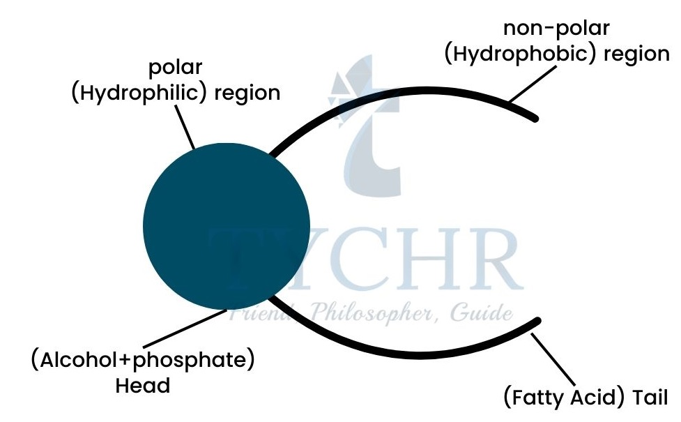
It is a continuous and constituent structure of phospholipids and the embedded proteins. In 1972, Seymour J. Singer and Garth L. Nicolson proposed a model of cell membrane named as Singer-Nicolson model also called the fluid mosaic model.
Phospholipid bilayer
- Phospholipid is made up of glycerol (3-C compound), which further includes fatty acids and alcohol group with phosphate.
- Two distinctive areas of bilayer are termed as hydrophilic (water-loving) and hydrophobic (water-fearing)
- Cholesterol molecules are not present in the plant cells but in animal cells.

Membrane Proteins

- Proteins in the cell membrane create the mosaic effect.
- They are very diverse in structure and their position in the membrane.
- Two types are found; integral and peripheral proteins.
- Integral proteins show an amphoteric nature, with both hydrophobic and hydrophilic regions within it.
- Peripheral proteins do not protrude into the middle hydrophobic region but remain on the surface only.
- Functions they perform:
- Acts as hormone-binding sites
- Impart cell-to-cell communication
- Useful in cell adhesion
- Catalyse enzymatic reactions
- Acts as channels for passive as well as active transport
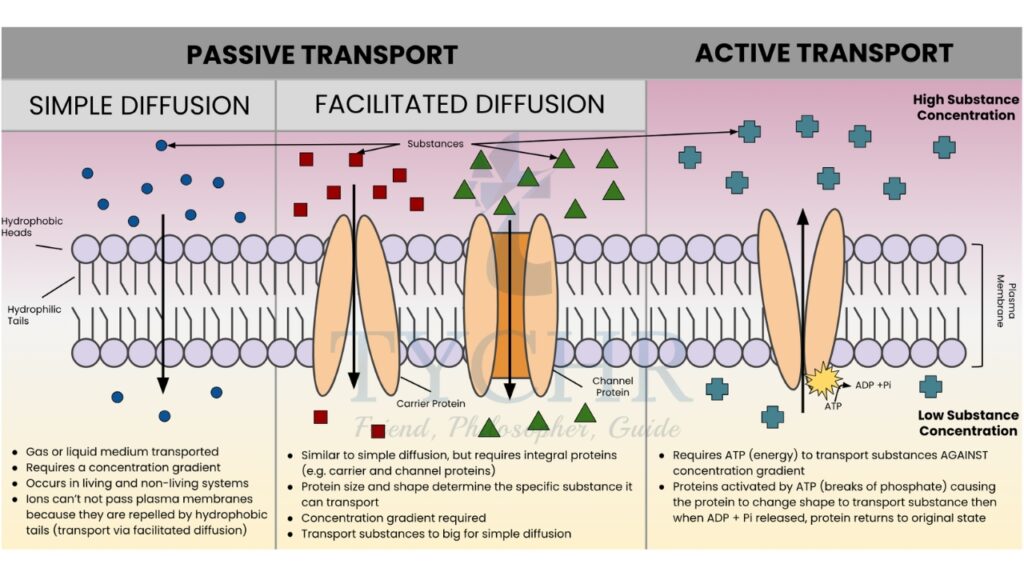
Membrane transport
Two types of cellular transport are found; passive and active transport.
I. Passive transport
- Does not require energy.
- Concentration gradient causes passive transport. Movement of substance occurs from an area of higher concentration to lower concentration.
- The movement of the substance will continue until the equilibrium is not reached. Once it’s reached, there will be no more concentration gradient and hence the movement from higher to lower stops.
- Diffusion: Movement of substance that occurs from an area of higher concentration to lower concentration across a membrane(partially permeable).
- Facilitated diffusion: Involves the membrane bound proteins to aid movement of substance across the membrane. The protein acts as a “carrier protein”.
- Osmosis: It involves the movement across the partially permeable membrane due to concentration gradient.
- Partially permeable membrane allows only certain molecules to pass through but not all.
- A solution with higher concentration of solutes is called hypertonic and with the lower concentration is called hypotonic. When there obtains the equal concentration on either side of the membrane (inner and outer), then it is called isotonic.
- Diffusion of the molecules depends on the size and the charge contained by them. Small and nonpolar molecules (gases such as O2, CO2, etc.) cross membranes easily but it is difficult for large and polar molecules (ions like K+, Na+, Cl+, etc.).
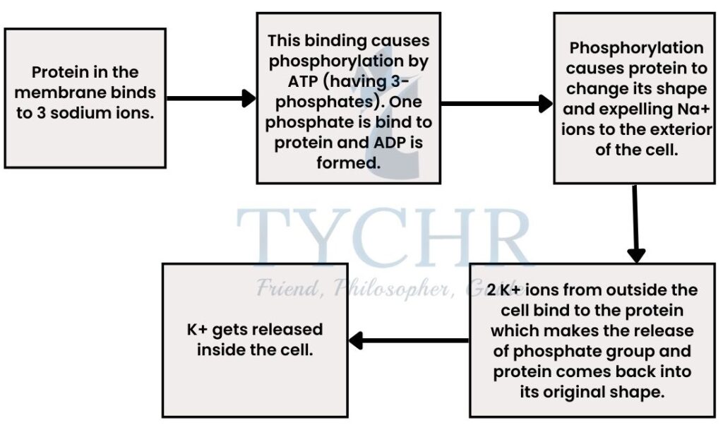
II. Active transport
- This transportation of molecules or substances requires energy which is provided in the form of ATP, because movement in this is against the concentration gradient.
- Sodium-potassium pump is the best example of active transport. Animal cells have higher concentration of K+ ions inside and higher concentration of Na+ ions outside the cell.
- Endocytosis and exocytosis
Endocytosis allows the molecules to enter the cell while exocytosis allows them to leave the cell. Fluidity (provided by bilayer structure) of the membrane is essential for these processes.


Cell division
Process of cell division
Cell division occurs as the cell cycle completes.
The cell cycle describes the growth and division phase of the cells. It occurs mainly in 2 parts or phases; Interphase and M phase (mitosis).
Interphase

- Longest phase of the cell cycle includes G1, S and G2 phases.
- G1 phase – 1st phase of cell growth in which it prepares for the next steps to come.
- S phase – (synthesis phase) Replication of DNA occurs.
- G2 phase – Cell grows and prepares for mitosis, DNA begins to condense, and microtubules may form.
- G0 phase – Resting phase, cell remains inactive.
Cyclins mediate the cell cycle by binding to cyclin-dependent protein kinases (CDKs). These activated enzymes cause cell to move from G1 S G2 M-phase.
Mitosis (M phase)
- The replicated chromosomal DNA separates, moves to the opposite poles inside the cell and the cell cytoplasm divides into two with the identical genetic material.
- In each of the daughter cells, this process includes four phases; prophase, anaphase, metaphase and telophase.
Prophase

- Chromatin fibers condense even more to form chromosomes.
- Nuclear envelope disintegrates and nucleoli disappear.
- Mitotic spindles formed.
- Centrosomes move towards the opposite poles.
Metaphase

- Chromosomes move to the equator of the cell in the middle, referred to as the metaphase plate.
- Movement of chromosomes by microtubules attached to centromeres starts as the metaphase comes to its end.
Anaphase

- Shortest phase.
- Microtubules start to contract.
- Sister chromatids start to split and move towards the opposite poles.
Telophase

- Chromosomes reach the opposite poles.
- Nuclear membrane begins to re-form.
- Nucleoli reappear and the spindle fibers disappear.
After telophase, it performs cytokinesis.
Cytokinesis
The division of cytoplasm into two daughter cytoplasmic cells is called cytokinesis.
This process occurs differently in animal and plant cells. In animal cell an inward pinching of the fluid plasma membrane forms cleavage furrows. And in plant cells there is the formation of cell plate in the midway of two separating cells Both of these
formations results in two daughter cells.
Cancer
An uncontrolled division of cells causes a cluster of unwanted cells which leads to the formation of a tumor. Two types are found; primary tumor and secondary tumor.
- Primary tumor occurs at the site of cancer but the secondary tumor is a metastasis,
which spreads from the original location of cancer to different parts inside an organism.
How do cancerous cells form? - The cellular information for division of cells is contained in the genes. Mutation of genes
causes them to convert into oncogenes, which further converts the normal cell into a
cancer cell which then divides abnormally

Cells are the building blocks of all living organisms. This study note has covered the essential aspects of cell structure, function, and processes. Applying this knowledge to real-world examples and biological systems is vital for a deeper understanding. For personalized support in IB DP Biology, including cell biology and other topics, consider working with a tutor. TYCHR connects students with experienced IB Biology HL & SL tutors who can provide tailored assistance. A strong foundation in cell biology is essential for success in IB DP Biology.
