Module 1.1: Localization
What will you learn in this section
- Localization of function is the idea that every behaviour is associated with a specific brain region
- Brain structure
a) Cortex
b) Cerebellum
c) Limbic system
d) Brain stem. - Research supporting strict localization
- Research opposing the idea of strict localization
- Relativity of localization: the split-brain study
Brain structure
- The nervous system is a system of neurons— cells that perform the function of communication in the body. The central nervous system consists of the spinal cord and the brain
- The major parts of the human brain are the:
a) cortex
b) cerebellum
c) limbic system
d) brain stem.
a ) Cortex
- The cortex is the layer of neurons with a folded surface covering the brain on the outside. It is the largest part of the human brain associated with higher-order functions such as abstract thought or voluntary action. Evolutionarily, this part of the brain developed the latest.
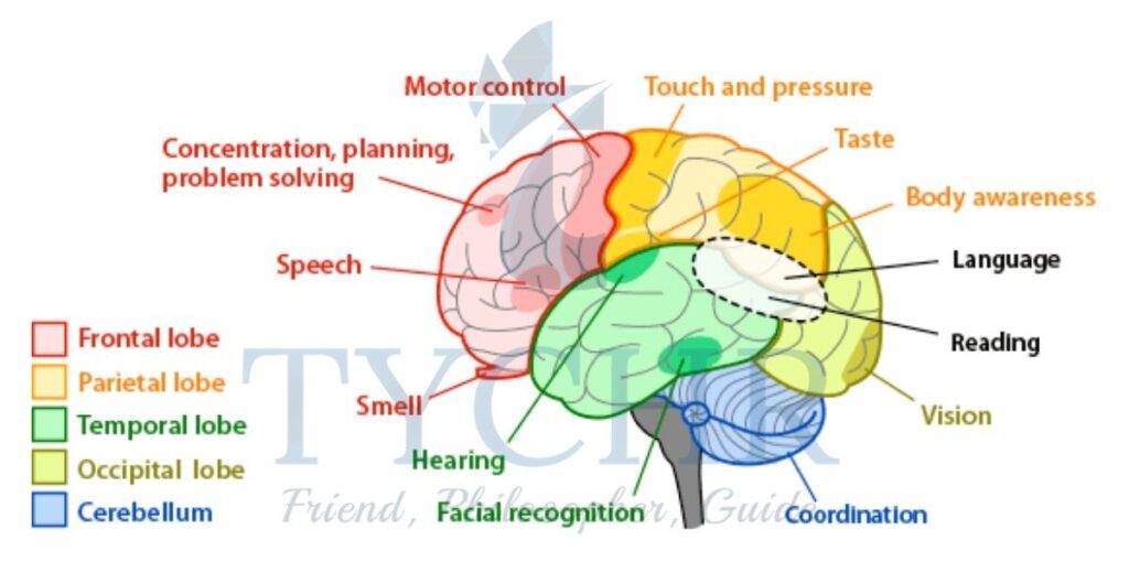
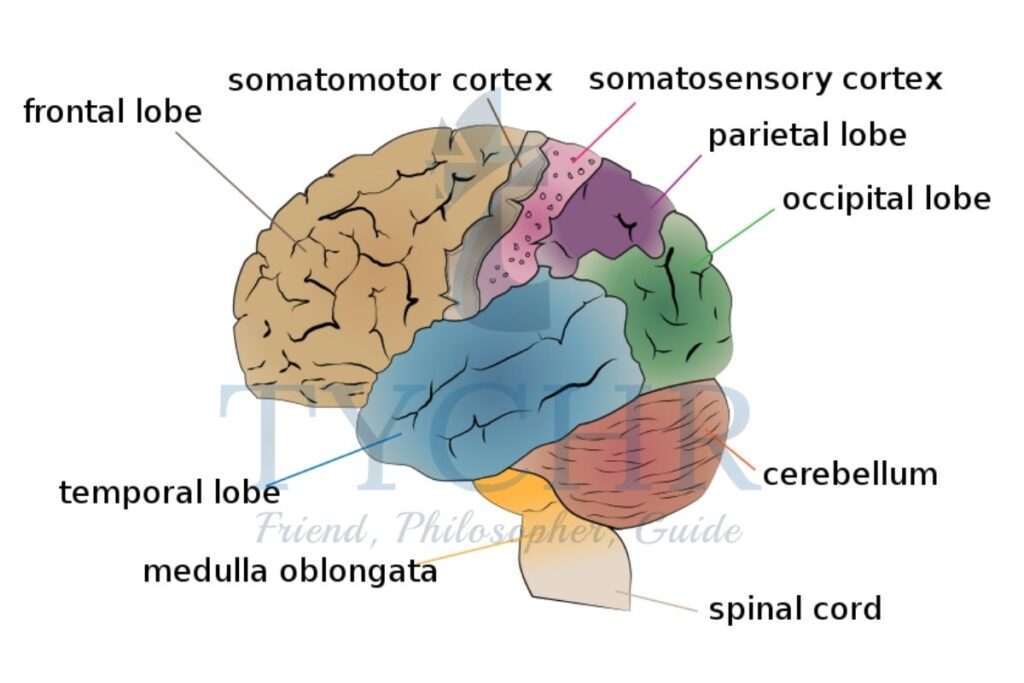
- The cortex is divided into four sections called “lobes” .
1. The frontal lobes are associated with reasoning, planning, thinking a nd decision-making, voluntary action, complex emotions, and so on.
2. The parietal lobe is associated with movement, orientation, perception and recognition.
3. The occipital lobe is associated with visual processing.
4. The temporal lobes are associated with processing auditory information, memory and speech. - There is a deep furrow along the cortex that divides it into the left and right hemispheres. A structure of neurons that connects these two hemispheres is known as the corpus callosum
b) Cerebellum
- The cerebellum (“the little brain”) got this name because it looks somewhat like the cortex: it has two hemispheres and a folded surface.
- It is associated with coordination of movement and balance.
c) Limbic System
- The limbic system is an evolutionarily older subcortical structure. It is sometimes referred to as the “emotional brain”. It includes several structures, some of which are as follows.
1. The thalamus has mostly sensory functions. Nerves from almost all sensory organs reach the thalamus as a final “hub” before they are connected to the cortex.
2. The hypothalamus is “below” the thalamus in the brain and it is involved in such functions as emotion, thirst and hunger.
3. The amygdala is involved in memory, emotion and fear.
4. The hippocampus is important for such functions as learning, memory and transferring short-term memory to a more permanent store, spatial orientation.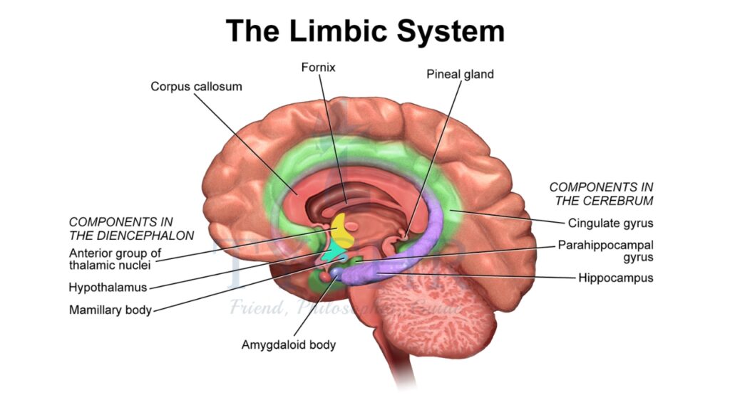
Localization
- Localization is the ability to identify parts of the brain which serve certain functions.
- The areas of brain which are of particular interest in terms of localization are
Hemispheric Localisation
- The brain is contralateral. This means that the right hemisphere relates to the left side of the body and the left hemisphere to the right side of the body.
- The right hemisphere has been found to have three key functions that have been supported experimentally: recognising emotion and faces, together with spatial functioning.
- Research {Dundas el: aI., 2015} supports the idea that the right hemisphere is dominant for recognizing faces as in research it has been shown that participants process faces predominantly in the right hemisphere. It has also been found that this recognition of faces is more strongly associated with the right hemisphere when the recognition is of very familiar faces [Bombari et al.2014}.
- However it should be noted that the brain becomes less lateralized with age with the non: dominant hemisphere supporting the superior hemisphere for any given task. This is called the HAROLD model {Hemispheric Asymmetry Reduction in Older Adults}. Therefore, in an experimental procedure it is important to consider the age of participants.
Motor centers
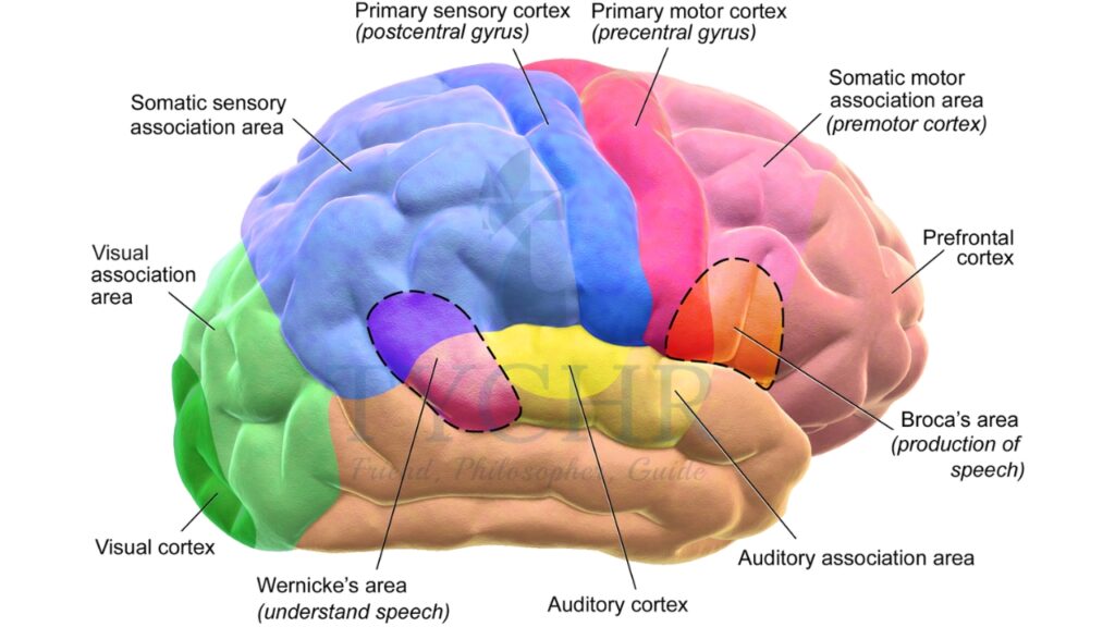
- The motor centers of the brain are concerned with movement. There are more simple movements like reflexes, coughing, gagging, sneezing, and so on: but the more complex movements we use are coordinated within the motor cortex, which can be found in the parietal lobe of the brain.
- Much of the motor cortex is actually concerned with small areas of the body such as the lips, tongue and hands, as these are areas that require fine motor movements to be performed.
Somatosensory cortex
- The somatosensory area of the brain is focused specifically upon sensory information. It is positioned next to the motor cortex in the parietal lobe and works closely with the motor cortex more to allow the individual to move appropriately around their environment.
Visual center
- The area of the brain which processes visual information is positioned in the occipital Lobe. It is called the primary visual cortex. There are actually two in the brain, one in both the left and right hemispheres. An area called Area VI within the primary visual cortex seems to be necessary for perceiving visual stimuli.
- This is apparent from cases where there has been damage in that specific area.
Auditory center
- There is a primary auditory cortex in both hemispheres. This is required to process complex sounds and is situated in the temporal lobe.
- If there is a lesion in the area then the individual can still hear some sound, but anything that is complex like speech or music cannot be processed.
- As the brain is contralateral, sounds heard by the right ear are processed predominantly by the left hemisphere and vice versa. However, unlike the motor cortex, there is not a clearly defined split as some sounds from the right car are processed on the right side of the brain too.
Language centers
- Our production and processing of language occur in many areas across the brain, depending on the function required and modality.r (ie. sound, written, oral production )of the language. This section considers two areas: Broca’s and Wernicke’s areas.
a) Broca’s area
- The function of Broca’s area is speech production and it is situated in the frontal lobe very close to the temporal lobe, in the dominant hemisphere.
- Ninety-six per cent of right handers and about 75 percent of left handers have the left hemisphere as their dominant hemisphere for language.
- Individuals with a lesion in this area cannot produce speech, but their understanding and processing of other aspects of language remain unimpaired.
b) Wernicke’s area
- Wernicke’s area is key to understanding language and, more specifically, speech. This area is close to the auditory cortex and can he found where the temporal and parietal lobes meet in the dominant hemisphere for language {which is usually the left hemisphere}
- It is linked to Broca’s area, as the two are closely related, with a bundle of connecting neurons called the arcuate fasciculus.
- Interestingly Wernicke’s area is also found in deaf people who use sign language which suggests that its purpose is for more than just speech.
- Individuals who have a lesion in this area are still able to produce fluent speech as Broca’s area is unaffected. although they will sometimes include words that have been made up. However, their ability to understand language is severely impaired so someone speaking to them will not understand both at sentence and word level.
Research restricting the idea of strict localization
- Karl Lashley (1890–1958) used the technique of measuring behavior before and after a specific seriously controlled induced brain damage the the rats’s cortex. In a typical study he would prepare the rat to run out a maze without errors in search of food. After learning occurred, he would remove an area from the cortex.
- Then he would place the rat back at the starting point of the maze and note down the change in behavior. He removed varying portions of the cortex in different rats, ranging from 10% to 50%. The main idea behind this was that if somewhere the memory of the maze is localized, then by removing area after area you can pinpoint towards the specific region in the cortex which is responsible for it. This search turned out to be a failure, so Lashley abandoned his own initial hypothesis. He concluded that memory was distributed rather than localized; a conclusion supported by the following observations.
- The principle of mass action based on a similarity observed between the percentage of cortex removed and the learning abilities. The lesser the cortex, the slower and more inefficient learning.. The main aim here is deterioration in performance depending on the cortex’s percentage destroyed but not in the area of the cells which are destroyed.
- Equipotentiality—This describes the capacity of one region of the cortex to adopt the functions of another.
- These observations led Lashley to conclude that memory is widely distributed across the cortex. This conclusion is mostly held up today. However, it has been proven that memory is not as same (and However, it has been shown that memory is not as evenly ( and similarly) distributed in the cortex as what Lashley expected.
- To some extent the difference in the two extreme positions (the localizationism of Broca, Wernicke and Pen eld versus the holism of Lashley) may be explained by the methods they used in their studies. Aphasia was being relied on by localisationists leading to brain damage. Holists looked into maze running behavior. However, learning to run through a maze is in itself a highly complex behavior that involves motor and sensory functions, so it may not be suitable enough for the study of localization.
- There needs to be a converging position that is a more accurate rejection of localization in the brain. Currently,: localization is supported by neuroscience by admitting localization for some functions under certain conditions, but it outlines the limits of localization clearly.
- Before we formulate these limits let’s look at another research study that demonstrates relativity of localization of function (that is, localization and distribution at the same time): split-brain research by Gazzaniga (1967) and Sperry (1968).
Relativity of localization: the split-brain research
- It has to be noted that split-brain studies represent research into lateralization—the division of functions between the two hemispheres of the cortex. Lateralization is a special case of localization.
- Research in this area was pioneered by Roger Sperry. Initially, the studies were conducted with animals, for example, cats.
- An opportunity to replicate the studies with humans emerged when it was discovered that surgically cutting corpus callosum was an effective measure against severe epilepsy with uncontrollable seizures. Roger Sperry was joined by Michael Gazzaniga, and in 1967 Gazzaniga published results of the research with human split-brain patients. Four of the ten patients who had undergone this surgical procedure by that time agreed to participate. The patients were examined thoroughly over a long time period with various tests.
- The aims of the study were to test the theory of lateralization and to see if the two hemispheres have uniquely different functions.
- Initial observations showed that patients seemed to be remarkably unaffected by the surgery. There was no change in their personality and intelligence, and one of the patients on awakening from the surgery joked that he had a “splitting headache” and recited a tongue-twister.
- The authors devised a technique where the participant had to sit in front of a board and look at the dot in the middle of it. Visual stimuli would then be presented for one tenth of a second either to the left or the right visual eld (the far left or far right on the board). Optic nerves from the left eye are connected in our brain to the right hemisphere, and vice versa. So, by presenting the stimulus to the left visual field the researcher “sends it” to the right hemisphere, and stimuli from the right visual field goes to the left hemisphere. Also, a variety of objects were placed behind the screen so that participants could feel them with their hands.
Module 1.2: Neuroplasticity
What will you learn in this section
- Definitions
- Neuroplasticity is the ability of the brain to change through the making and breaking of synaptic connections between neurons; causing factors are both genetic and environmental
- Different scales of neuroplasticity—from synaptic plasticity to cortical remapping
- Remapping of the sensory cortex
- Merzenich et al (1984): cortical remapping of sensory inputs from the hand occurs within 62 days in owl monkeys— adjacent areas spread and occupy parts of the now unused area for the amputated digit
- Neuroplasticity as a mechanism of learning
- Example 1—Draganski et al (2004): learning a simple juggling routine increases the volume of grey matter in the mid- temporal area in both hemispheres; lack of practice makes this area shrink, but not to the original size
- Example 2—Draganski et al (2006): learning large amounts of abstract material leads to an increase of grey matter in the parietal cortex and the posterior hippocampus
Definitions
- Neuroplasticity is the ability of the brain to change throughout the course of life. When the synaptic connections are made and broken a change occurs between the neurons. In this process neural networks in the brain literally change their shape.
- Genetics(generally pre programmed development of the brain) and environmental (for eg: injury or damage in the brain or simply learning new skills) are responsible.
- Neuroplasticity can be observed on different scales.
a) On the smallest scale, at the level of a single neuron, it takes the form of synaptic plasticity: the capability of the neuron to generate new synaptic connections and break the old ones
b) On the largest scale, neuroplasticity takes the form of cortical remapping: the occurrence of the brain area X assumes brain area Y’s function for example, due to injury. - Synaptic plasticity depends on the activity of neurons. If two close-by neurons are oftenly activated at the same time; a synaptic connection between the two neurons may slowly form. Likewise if two neurons are hardly activated together the functioning connection may slowly fall apart. This has been summarized like this: “neurons that are together, wire together” (which was originally said by Carla Shatz and is quoted in Doidge, 2007) and “neurons that are out of sync, fail to link” (Doidge, 2007, pp 63–64).
Remapping of the sensory cortex
- One of the early studies of neuroplasticity on the level of cortical remapping was done by Merzenich et al (1984). Researchers studied the cortical representation of the hand in eight adult owl monkeys. The procedure involved three steps.
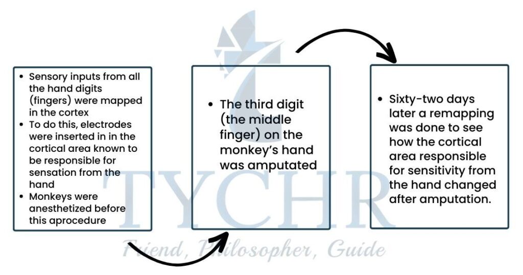
- Results of the first mapping showed that there were five distinct areas in the brain, each responsible for one finger, and adjacent fingers were represented in adjacent areas in the cortex. It was found that the adjoining area (the ones responsible for sensitivity from digits 2 and 4) spread and occupied parts of the area which is unused. The areas responsible for digits 2 and 4 became larger while the areas responsible for digits 1 and 5 stayed the same. It was concluded that cortical remapping of sensory inputs from the hand occurs within 62 days in owl monkeys.
Neuroplasticity as a mechanism of learning
- Example 1—Draganski et al (2004): learning a simple juggling routine increases the volume of gray matter in the mid- temporal area in both hemispheres; lack of practice makes this area shrink, but not to the original size.
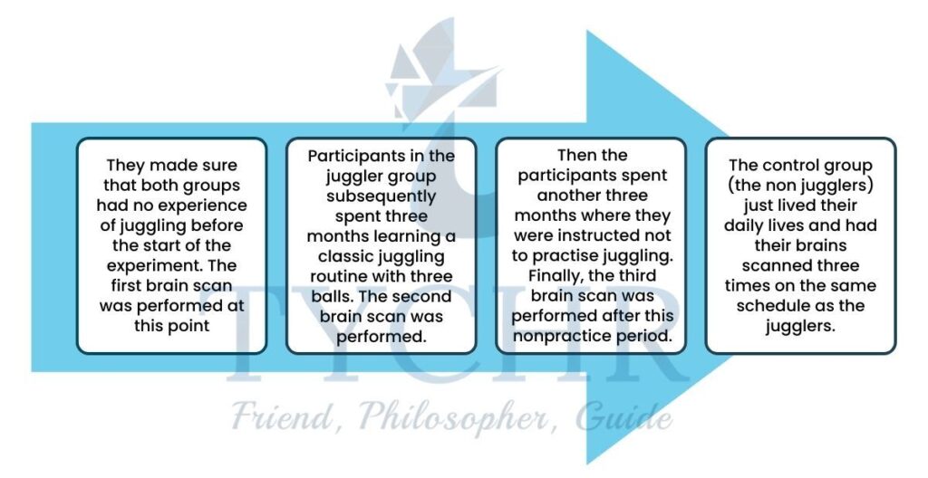
- The researchers used a random sampling design and a self-selected sample—they randomly allocated a sample of volunteers into one of two groups: jugglers and non-jugglers
- No difference was shown in the brain structure in the scans of the two groups before the start of the experiment
- At the second scan, however, more gray matter was seen in some areas of the cortex of the juggler group , the mid temporal in both the hemispheres. These areas were known to be implicated in coordination of movement.
- These differences got smaller on the third scan, but there was still more gray matter in these areas in jugglers than there was on the first scan. Also, a correlation was seen between the performance of the juggling and amount of change brain changes in participant When you fail to practice, they shrink back significantly (perhaps not to the initial state, though)
Example 2 — Draganski et al (2006):increasing the amount of gray matter in the parietal cortex and posterior hippocampus by studying a lot of abstract material.
- Draganski et al (2006) glanced at 12 subjects of control and about 38 students studying medical matched for the age and gender.
1. The first scan was obtained three months before the examination,
2. The second scan on the first or second day after the examination, and
3. The third scan three months later (after the examination the students had a break). - Results showed that, although there were no differences in regional gray matter at baseline, there were two major changes occurring in the brains of the medical students.
a) The gray matter was increased in the parietal cortex in both the hemispheres respectively. By the time of the third scan The volume of gray matter in this region did not reduce. Studying for an examination in medicine has a more lasting impact on the brain. The changes stay with you even after a study break.
b) The gray matter in the posterior hippocampus grew. The pattern was different here. The gray matter was slowly increased comparative to the first scan in the third. That is, gray matter in the hippocampus continued to grow even after the examination.
c) Stress is known to reduce gray matter volume in hippocampal regions. This may have resulted in two opposite effects on the hippocampus simultaneously between the first and the second scan: learning increased gray matter in the posterior hippocampus but examination stress decreased it. After the examination this negative influence of the examination stress was corrected and the lost hippocampal volume was restored, while the gray matter that was formed due to learning remained
Module 1.3: Neurotransmitters
What will you learn in this section
- Nervous system processes
- The structure of a neuron
- Electrical processes: threshold of excitation, action potential
- Chemical processes: neurotransmitters and how they function
- Excitatory and inhibitory neurotransmitters
- Agonists and antagonists
- Limitations in neurotransmitter research
- Effect of serotonin on prosocial behaviour
Serotonin reduces acceptability of personal harm and in this way promotes prosocial behaviour Crockett et al (2010)—participants solved moral dilemmas after receiving a dose of either citalopram (an SSRI) or placebo - The role of serotonin in depression
The serotonin hypothesis: low levels of serotonin in the brain play a causal role in developing depression - Effect of dopamine on romantic love
Fisher, Aron and Brown (2005): looking at pictures of loved ones is associated with higher activity in the dopaminergic pathway—a system that generates and transmits dopamine and increases dopamine-related activity in the brain - The role of dopamine in Parkinson’s disease
Freed et al (2001): transplantation of dopamine-producing neurons in the putamen of patients with severe Parkinson’s disease results in some clinical benefit in younger but not older patients
Nervous system processes
The structure of a neuron
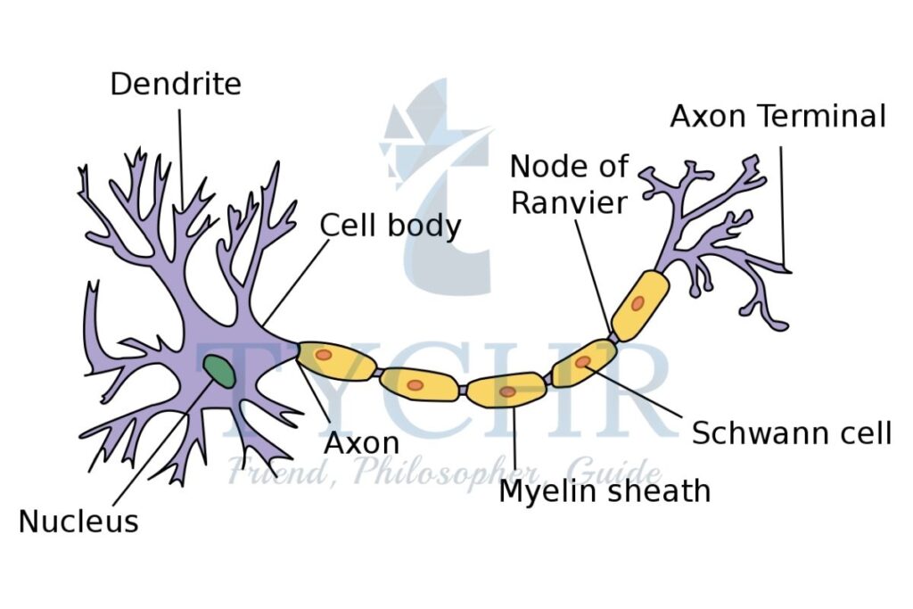
- A neuron consists of three parts:
- the body (soma),
- dendrites and
- axon.
- Dendrites and axon are filaments that extrude from the soma: typically multiple dendrites but always a single axon. Dendrites and soma are responsible for receiving signals from other neurons,
- where as axons are responsible for further transmitting signals. Where the axon of one neuron approaches a dendrite or soma of another neuron, a synapse is formed. This means that a synapse (or a synaptic gap) is a structure that connects two neurons: the word “synapse” comes from the Greek synapsis meaning “conjunction”.
Electrical ( threshold of excitation, action potential) and Chemical processes (neurotransmitters and how they function)
- The nature of information transmission in the nervous system is partly electrical and partly chemical.
- Every neuron has a certain threshold of excitation received from the other neurons, and if the sum excitation exceeds this threshold, the neuron “fires”—generates a brief pulse called action potential that travels with the axon to the remaining neurons, passing the excitation further.
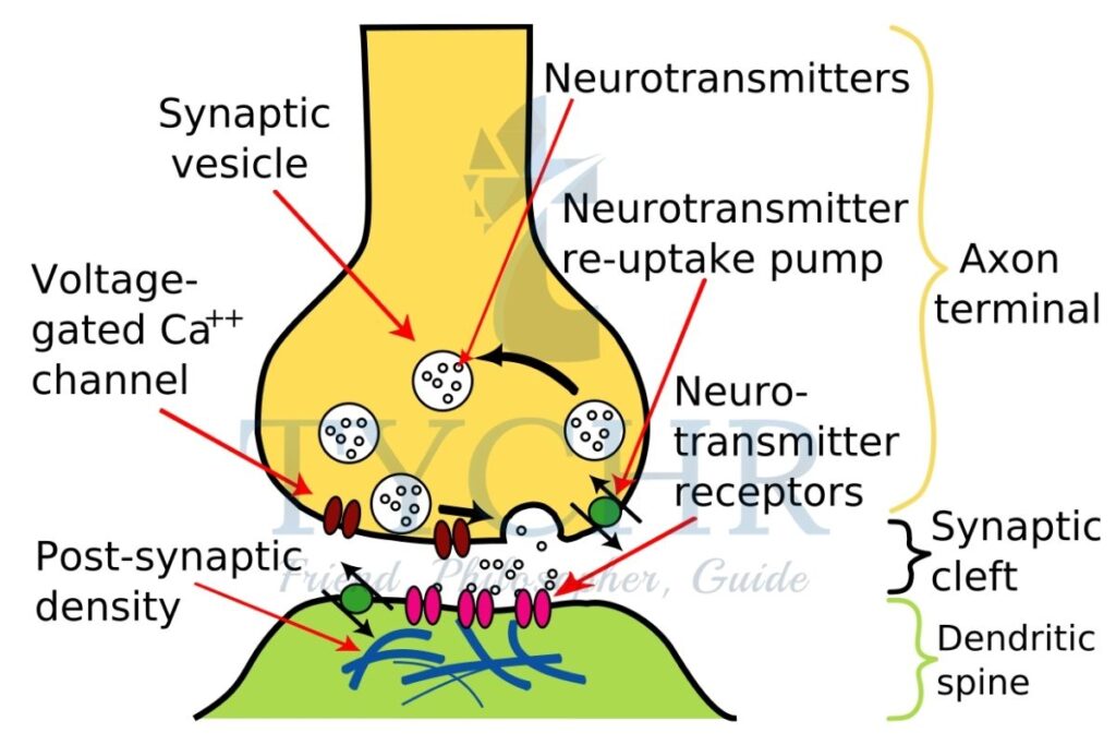
- The pulse reaches the end of the axon and there, the transmission of the mechanism becomes chemical at the gap of synaptic.
- This happens as follows.
- When the action potential stretches to the very end of the axon, neurotransmitter is let out from the terminal of the axon into the gap of synaptic.
- Neurotransmitters are chemical messengers. They are constantly synthesized in the neuron and moved to the axon terminal to be stored there. For a short period of time in the gap of synaptic a let out neurotransmitter is available whilst it may be destroyed (metabolized), and made to return into the pre synaptic axon terminal by reuptake or reach the post- synaptic membrane and bind to one of the receptors on its surface.
- If the neurotransmitter binds to a receptor in the postsynaptic membrane, this process makes a difference in the membrane potential and so it provides aid in activating an electric pulse in the postsynaptic neuron. Here the chemical mechanism becomes electrical again.
Neurotransmitters
- Dendrites and soma are liable for getting signals from different neurons, though axons are answerable for additional communicating signals.
- Excitatory :- To cross the synapse, excitatory neurotransmitters grant permission. The brain is stimulated by their effects.
- Inhibitory:- The impulse is halted by inhibitory neurotransmitters, which prevent it from traveling through the synapse. The brain is calmed by their effects.-
- These neurotransmitters are always in a state of dynamic balance. When excitatory or inhibitory neurotransmitters are not within the optimal ranges in the brain it may lead to various behavioral malfunctions such as mental disorders.
Agonists and Antagonists
- Neurotransmitters themselves are affected by
- Agonists :Agonists are synthetic compounds that upgrade the activity of a synapse-
- Antagonists:- Chemicals called antagonists work against a neurotransmitter to prevent the signal from being transmitted ahead.
- Many drugs function as agonists or antagonists. For example, a class of drugs known as SSRIs (selective serotonin reuptake inhibitors) selectively inhibit (block) the reuptake of the neurotransmitter serotonin from the synaptic gap. This increases the concentration of serotonin in the synapse. SSRIs have been shown to be effective against depression.
Limitations in neurotransmitter research
- As you see, neurotransmission is a complex process determined simultaneously by multiple factors.
- Imagine we have artificially increased the level of neurotransmitter X in the brain and this resulted in a change of behavior Z (for example, elevated mood). Can we say that neurotransmitter X influences elevated mood? Yes, to a certain extent, but with a lot of limitations to be kept in mind.
- X may function as an agonist for neurotransmitter Y, which in turn may affect behaviour Z. In other words, the effects of neurotransmitters may be indirect,
- X may serve as a trigger for a long-lasting process of change in a system of interconnected variables. In other words, the effects of X on Z may be postponed.
- X is usually not the only factor affecting Z.
- X is never the only factor that changes. As you artificially increase the level of X, this may result in various side effects.
- Research into the influence of neurotransmission on behavior will therefore always be reductionist in the sense that we need to manipulate one variable (X) and assume that it is the only variable that changes.
Serotonin
- Serotonin is an inhibitory neurotransmitter that is involved in sustaining stable mood and regulating sleep cycles, for instance.
- It is found in the gastrointestinal tract as well as the brain and is implicated in mood regulation.
- Changes in the levels within the brain are evident when an individual is experiencing an elevated mood and lower levels are observed when there is a depressed mood.
- The recreational drug ecstasy raises levels of serotonin and its ameliorating effect on mood is well researched. Low levels of serotonin are associated with depression and SSRls {selective serotonin uptake inhibitors}. which are antidepressants that raise the levels of serotonin within the brain and help some patients suffering from depression.
- Another potential effect of serotonin levels on behavior is aggression. Increased levels of aggression when serotonin levels are lower have been found in humans This is thought to occur through the serotonin levels affecting emotion processing in the brain.
Dopamine
- Dopaminergic pathway is a system that generates and transmits dopamine and increases dopamine-related activity in the brain. It is a reward system because dopaminergic activity is associated with motivation and feelings of pleasure.
- The ventral tegmental area (VTA) and caudate nucleus, both these regions are rich in dopamine and form the key part of the so-called dopaminergic pathway.

Role of Dopamine in Parkison’s disease
- Parkinson’s disease is a degenerative disorder that mainly affects the motor functions of the nervous system. The early symptoms of the disease are shaking, rigidity, and difficulty with movement and walking. Later in the development of the disease, thinking and behavioral problems also occur. For now Parkinson’s disease doesn’t have a cure and the exact causes are unknown
- The dopamine hypothesis focuses on the idea that low levels of dopamine in the system of an
- individuals are linked to the onset of parkinson’s while high levels of dopamine in the system of an individual are linked to the onset of schizophrenia. High levels of dopamine appear to increase the activity within the neurons, which means the level of communication is also increased.
Module 1.4: Hormones and Pheromones
What will you learn in this section
- Difference between hormones and neurotransmitters
- The function of hormones
- Examples of hormones
- Examples of phermones
Difference between hormones and neurotransmitters
- In their function hormones are similar to neurotransmitters: essentially Both are chemical messengers. However, neural communication (neurotransmission) and hormonal communication differ in a number of ways.
- Hormones are released into the bloodstream and travel with blood to reach their destination. Conversely, with the nervous cells neurotransmission is the communication. Because of this the hormones cover the areas where the nervous system cannot, as the blood vessels network is more elaborated The nervous system supervises relatively fast process (movement, emotion, decisions, and so on), whereas hormones are able to regulate the enduring ongoing processes like metabolism , growth, reproduction and digestion.
- Normally, the voluntary control over neural regulation is higher comparative to the hormonal regulation. For example, it is possible for you to control your emotions to a certain extent, whereas the degree of control you have over your growth is negligible.
- However, it should be noted that the nervous system and the endocrine system are interdependent. These two systems interact and to some extent can influence each other. Also, some chemicals at times could be both neurotransmitters and hormones for example, adrenaline.
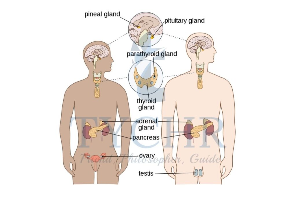
- Hormones are released by endocrine glands: adrenal glands, hypothalamus, pineal gland, pituitary gland, thyroid, parathyroid, thymus, pancreas, testes and ovaries. Together, these form the endocrine system.
Hormones
- Cells can be only influenced by hormones if they have the receptors of the hormone respectively. These cells are known as target cells. When a particular hormone ties to a receptor it releases a sequence of changes, and in that some of them are genomic, meaning gene activation or gene suppression. Most importantly this means that the hormones do not have an influence on the behavior directly. Instead, they take a turn to the probability that a particular behavior is bound to occur in response to a particular environmental stimulus. This is like buying ice-cream on a hot day: hot weather itself does not cause you to buy ice-cream, but it certainly increases the probability that you will.
- There are a variety of hormones produced in the body and they all have different functions. The most well-known hormones include adrenaline, noradrenaline, cortisol, oxytocin, insulin, testosterone and estrogen.
1. Oxy-tocin
It is mainly produced and released in the blood that is the pituitary gland. hypothalamus and released into the blood by the pituitary gland. It plays a vital role in the process of sex reproduction , childbirth and social bonding of a human being. It has been referred to as “the love hormone”, “the bonding hormone” and “the cuddle chemical”. During wet nursing, for instance, stimulation of the nipple releases oxytocin, which helps to improve the relationship between the mother and her child. A person also releases oxytocin when they hug or kiss another person.
2. Testosterone
- Testosterone is an androgen: a male hormone. It is not found exclusively in males but generally the amount found in females is much lower than that found in the male population.
- Testosterone is often linked to aggression and research has shown a link between the hormone and aggressive behavior. However, this appears to be predominantly in non-human animals.
- In humans, the relationship is more complex1 with testosterone not being implicated in all aggression, but mainly aggressive reactions to social provocation
- It is also implicated in behavior classified as masculine, as it is an androgen.
3. Adrenaline
- The heightened state brought about by an adrenaline increase is well-known. The term ‘adrenaline rush’ is widely used for the feeling that an arousing situation can bring about, and the description ‘adrenaline junkie’ is used to describe someone who actively seeks an adrenaline rush.
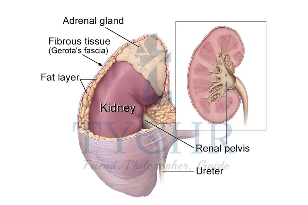
- Adrenaline is secreted by the adrenal glands located on top of each kidney. When an individual perceives a threat or potentially stressful situation, adrenaline rushes through the system triggering a racing heart and tense muscles, along with other effects.
- All these are part of the flight response. The term fight or flight’ is used to
describe the circumstances under which adrenaline is released into the body; this response will probably save everybody’s life at some point. It is designed to allow the body to deal with threat and danger. - While encountering the battle or a flight reaction, an individual can move quicker [due to the strained muscles] and take in more oxygen {due to the respiratory changes}. This enables a rapid
exit from the stressful situation, or indeed the ability to fight. The raised heartbeat helps to send the adrenaline around the system and gets blood to the muscles and organs. This means that a relationship between the physiological and behavioral reaction to situations and adrenaline is likely. - High levels of both adrenaline and testosterone have been found in a personality type called ‘sensation seeking’. People who are sensation seekers are able to cope with and like high levels of stimulation in the environment because they like their body to be in a heightened state of
arousal such as riding roller coasters, parachuting and car racing .
Pheromones
- The word pheromone is derived from a Greek word “phero” which means I carry and hormone which means stimulating, so pheromones are chemicals that “carry stimulation”.
- These chemical substances that are emitted by humans and nonhuman animals into their environment. This can occur in several ways, but the most frequent release of pheromones is in the sweat of the individual. These chemical substances are thought to elicit an effect on the environment itself and other animals in the environment.
- In non-human animals pheromones are a method of communication. Transmission occurs via sweat and other ۥ fluids such as urine. Humans are also thought to be affected by pheromones. Those argued to elicit a specific behavioral effect are called releaser pheromones and in certain contexts or situations, they trigger specific behavior. In other situations, the same pheromone may act as more of a primer.
- An example of the distinction between these two studies’ effects is found in the pheromones in male mouse urine. The releaser effect role is to make a person’s behavior more aggressive when a meddler is perceived. Menstrual synchrony is also promoted by the pheromone in the female mice.
Localization of processing pheromonal information in the brain
- Pheromonal information in animals’ brains is not processed in the same brain regions as ordinary smells, despite the fact that many of the pheromones can smell. The main olfactory bulb refers to the part of the brain that processes smell. Pheromones, on the other hand, are processed in a different way than other smells.
- The vomeronasal organ (VNO) is a separate structure found in mammals’ anterior nasal cavities. In the brains of animals, nerves from the VNO connect to the accessory olfactory bulb. According to Hertz (2009), this area is separate from the main olfactory bulb but adjacent to it.
- Due to the fact that humans lack either the VNO or the accessory olfactory bulb, it can be challenging to extrapolate animal research to human behavior. However, we need to be accurate on this point: The accessory olfactory bulb is present in human fetuses, but it regresses and disappears after birth. The VNO is present in some people, while others do not. Even in individuals who have the VNO, it appears to be inoperable: There is no connection to the brain or spinal cord. If the human brain actually processes pheromonal information, it must be processed elsewhere.
Research into attraction
- Pheromones are implicated in attraction in both humans and nonhuman animals. There are specific pheromones secreted that arouse and encourage sexual activity in males and females. In the animal kingdom. The boar pheromone is secreted via the male boar’s saliva when he is sexually aroused and this transfers to the air. The sow will detect the pheromone nasally and assume a standing position that makes it possible for the boat to mount
- In humans, there is thought to be an effect from secretion of a steroidal compound found in male sweat called androstenedione. It is found in women too, in lower levels. They found that the literature indicates that exposure to androstenedione does indeed increase attractiveness in mates, improves mood and also increases sexual amuse and desire.
Research into menstrual effects
- Stern and Me Clintoelr. [1998) found that when women received ‘odorless compounds from women’s armpits in the latter half of their menstrual cycle, their menstrual cycle was shortened, presumably by the effect of the other women’s pheromones as they approached the end of their cycle.
- The compounds were transferred hv the women wiping a pad, which had previously been wiped across the donor’s armpit, above their upper lip.
- However, if the compounds {which included pheromones } were collected from women at the beginning of their cycle, this had the opposite effect, lengthening the cycle of those who had received the compound. This shows that the menstrual cycle of a woman can be altered by communication via pheromones. This is a priming effect.
Module 1.5: Genes and behavior; genetic similarities
What will you learn in this section
- Genotype and Phenotype
- Genetic similarities: Twin Study
- The Influence of genetics on the environment
- Aggression and genetics
- Anxiety and genetics
Genotype and Phenotype
- All cells in the human body that have a nucleus contain a set of chromosomes .
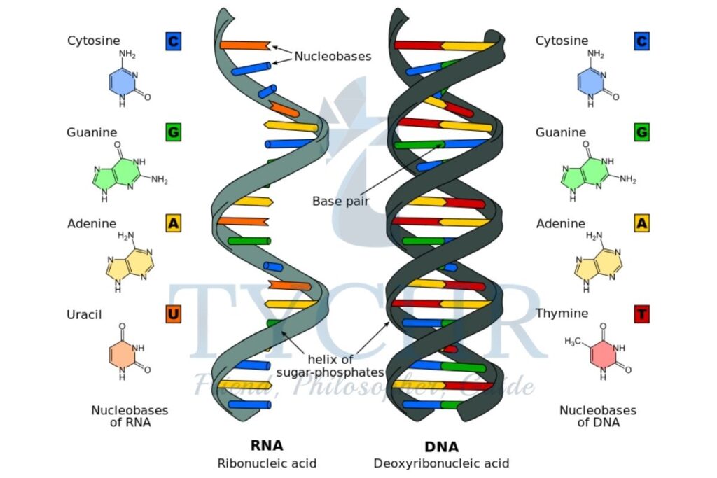
- A chromosome is a thread-like structure that contains a DNA molecule. The long DNA molecule is tightly coiled many times around supporting proteins, so a chromosome is a “package” that contains folded DNA.
- DNA (deoxyribonucleic acid) stores information. It is a code made up of a long sequence of four chemical bases (A = adenine, G = guanine, C = cytosine, T = thymine). The bases are paired up, making a sequence of base pairs.
- The DNA has a characteristic structure of the double helix which looks a bit like a ladder where base pairs are the ladder’s rungs (US National Library of Medicine, 2017). Information is coded in this sequence of bases like letters in a sentence (change the order of letters and you get a different sentence).
- The long sequence of chemical bases is broken up into 23 chromosomes, so each chromosome contains a part of the sequence. Each chromosome is present twice in each cell (except for sex cells).
- Humans have 23 pairs of chromosomes. One of the chromosomes in Each pair is from your mother and the other one from your father. Both the chromosomes in the pair have a code for identical characteristics (height, eye color, and so on), but the chromosomes themselves might not be identical.
Chromosome
- If DNA is one extremely long sentence, and base pairs are letters, then genes are probably words. A gene is a unit of heredity, a region of DNA that encodes a specific trait or function.
- For example, there is a gene for eye color, a gene for height, and so on. The total number of genes in the human organism is currently estimated to be around 20,000.
- Alleles are different forms of the gene. They can be dominant or recessive. The trait controlled by the recessive allele only develops if the allele is present in both chromosomes in the pair, whereas the trait controlled by the dominant allele will develop if at least one of the chromosomes in the pair contains it.
- For example, in the gene that codes for eye color the allele for brown eyes is dominant and the allele for blue eyes is recessive. So you will have blue eyes only if both the alleles in your chromosome pair are recessive.
- The set of traits as coded in an individual’s DNA is called genotype. The set of traits that actually manifest in an individual’s body, appearance or behavior is called phenotype.
- Phenotype comprises observable characteristics (eye color, height, and so on) and unobservable characteristics (blood type, immune system, and so on), as well as behavior.
- Genotype is the “plan” and phenotype is its implementation.
The Nature –Nurture Debate
- Nature-nurture is the long-lasting debate in psychology and philosophy that attempts to establish whether human behavior is determined primarily by biological factors such as genetics and brain structure (that is, nature) or environmental factors such as education and friends (that is, nurture).
Genetic similarities: Twin Study

- One of the key ways that genetic research can be conducted is by using twin studies. By using twin pairings, the level of heritability for any given behavior can, in theory, be calculated due to the genetic relatedness of the individuals.
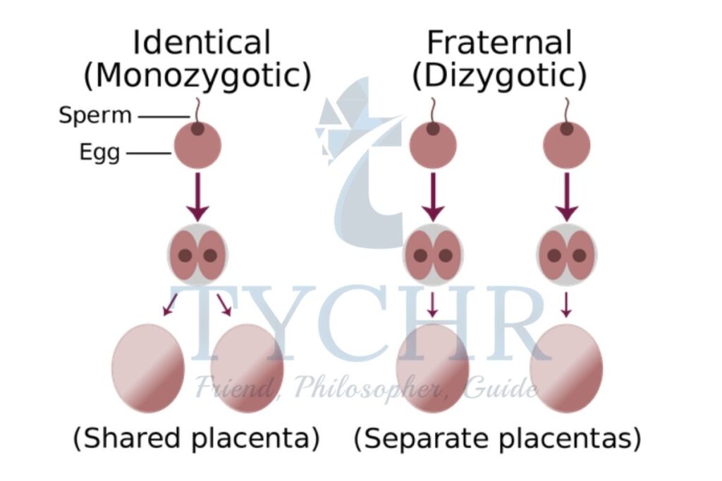
- There is a core assumption that underpins this type of methodology. As identical {monozygotic or M2} twins come from the same fertilized egg, they are 100 per cent genetically similar and non-identical i{dizygotic or DZ} twins share half their genes, as they come from different fertilized eggs, so any differences in behavior should be attributable to how similar the twin’s genetics are.
- This works on the assumption that the environment is the same for both twins {i.e. shared womb and home environment}. For example, if all the identical twins demonstrate a behavior and only half of the non-identical twins do so, then there is the suggestion that genetics can explain the behavior. In the real world, this rarely happens and concordance rates are used to ascertain the level of heritability.
- Concordance rates are expressed as a percentage. The percentage is calculated from the number of twin pairs, where both twins exhibit the behavior.
Adoption study - If identical twins have a concordance rate of 30 per cent for a behavior and non-identical twins have a 32 per cent concordance rate for the same behavior, the difference between the two concordance rates can be attributed to the differences between the two types of twins (M2 or D2} in terms of genetic similarity.
- However, it can also be said from the concordance rates that there are environmental influences because
otherwise the concordance rate for identical twins would be 100 percent. - Family studies are also conducted to examine behavior and its heritability level. By looking through a family tree and looking at members who may be diagnosed with a condition geneticists have estimated heritability of that condition.
The Influence of genetics on the environment
- Genes and environment are not completely independent: in many instances genes influence the environment too.
- One form of this dynamic development is niche-picking: the situation in which a person’s genetic predisposition causes them to choose environments that start to affect their behavior. For example, a child who is likely to be open to depression may deliberately seek high demanding environments where it is genuinely not easy to succeed. Niche-picking may explain one interesting property of heritability coefficients: they change during life, typically becoming larger. This means that if you use a sample of adolescent twins and the Falconer model to arrive at an estimate of heritability (A), this estimate will typically be smaller than if you use a sample of older twins. As you grow up, your genetic programme “unfolds’ ‘ causing you to choose certain “niches’ ‘ in the environment. In this way, in terms of their behavior, MZ twins become more and more similar with age.
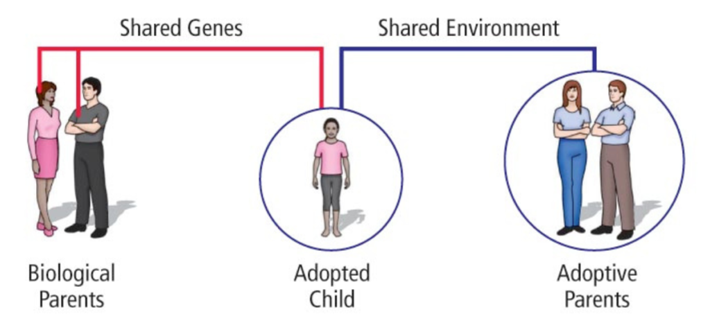
- Adoption studies provide a direct test of environmental malleability of cognitive abilities. There are two aspects of adoption studies that may provide slightly different angles on the nature nurture problem. These aspects are:
a) computing the correlation between cognitive abilities of the adopted child and the adoptive parents and comparing it to the correlation between cognitive abilities of the adopted child and the biological parents
b) comparing cognitive abilities of adopted children to those of their siblings who were not adopted but raised by their biological parents.
Aggression and Genetics
- The link between testosterone and aggression was discussed briefly in the previous section, It is also entirely possible that the high levels of testosterone implicated in aggression are a consequence of the individual’s genetic code.
- if this is indeed the case then it would be expected that the higher levels of testosterone would also be evident in the
individual’s relatives. Other bio-chemicals such as serotonin. dopamine and adrenaline are implicated too. - Genes can affect testosterone levels, as they have an effect on the way that the hormone is metabolized {this is the word used when a biochemical is processed by the body}.
- In terms of brain physiology, the frontal lobe is known to have properties that exert control and therefore some individuals’ brain physiology will mean that the control element is not as strong, leading to aggression.
- This frontal lobe physiology could be genetically determined. However, merely demonstrating how the genes could prompt changes to the ph biology and biochemical make-up of an individual is not sufficient; there needs to be an identifiable gene, or series of genes, if the genetic link to behavior can be made.
Anxiety and genetics
- Anxiety is a feeling of apprehension and worry. For many sufferers of anxiety disorders , anxiety is not enabling in any way, indeed it means that life can be difficult as the individual is held back by their anxiety.
- Evolutionary psychologists argue, however, that anxiety is evolutionarily adaptive. It prevents the person who is experiencing the anxiety from potentially putting themselves in danger, which increases their chances of survival.
- The problem comes when the anxiety levels become too high and are not adaptive. Nesse & Williams (1995) called the anxiety regulation mechanism the ‘smoke detector principle’.
- They argue that it is an evolutionary adaptation to have a heightened awareness of potential threats and that manifests itself as anxiety. However, Like a smoke detector, it is set to be triggered at the smallest sign of threat and, like the smoke detector, it is necessary for us to be aware of problems.
Module 1.6: Techniques used to study the brain in relation to behavior
What will you learn in this section
- Neuroimaging techniques
- Computerized axial tomography (CAT)
- Magnetic resonance imaging (MRI)
- Functional magnetic resonance imaging (fRMI)
- Positron emission tomography (PET)
- Electroencephalography (EEG)
- There are a variety of techniques used currently to investigate relationships between the brain and behavior, they are collectively known as brain imaging techniques, or neuroimaging.
These brain imaging techniques use varying ways to collect the information on brain structure and activation - Five of the most commonly used brain imaging techniques are:-
- computerized axial tomography (CAT)
- positron emission tomography (PET)
- magnetic resonance imaging (MRI)
- functional magnetic resonance imaging (fMRI)
- electroencephalography (EEG).
a) Computerized Axial Tomography (CAT)
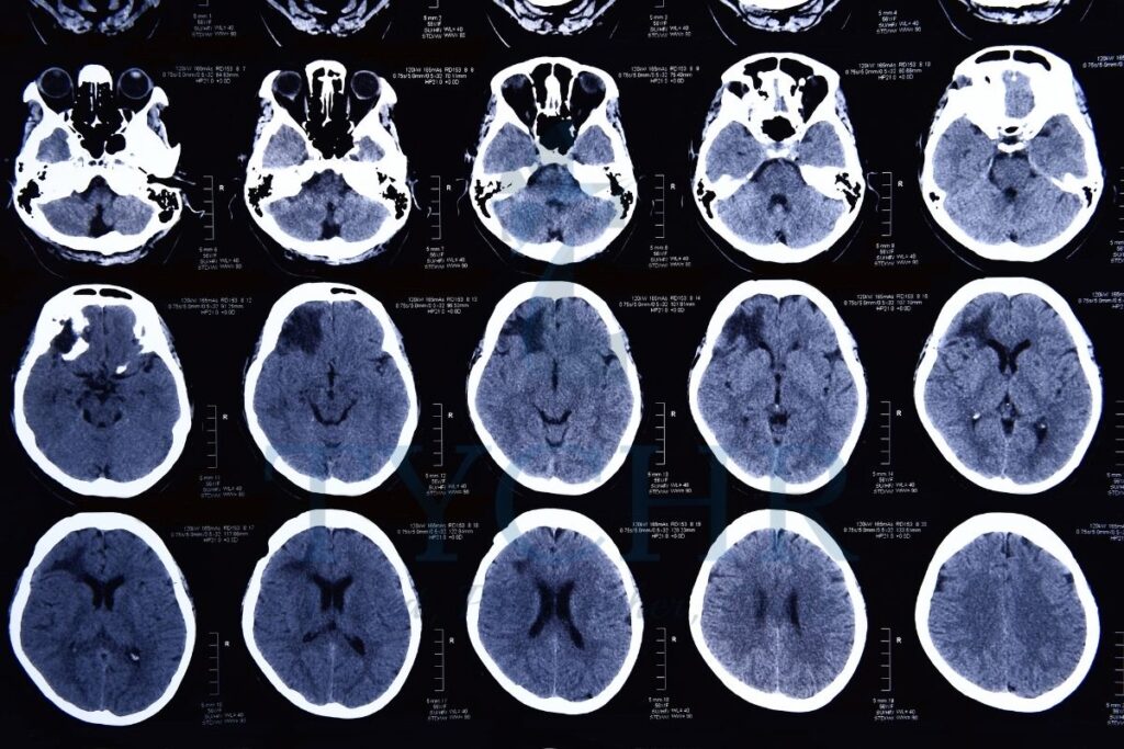
- Computerized axial tomography (CAT) works on the principle of differential absorption of X-rays. The object is on the table that slides inside a cylindrical apparatus, one-half of the cylinder projects X-rays across the cylinder to the other side, traveling through the brain. The other side of the cylinder has a detection unit to detect the X-rays. Using this movement across the front of one side of the cylinder to the other, many still photos can be taken as the cylinder rotates around the head. When all the images are merged they form a 3-D picture of the physiology of the brain.
- Bone and hard tissue absorb X-rays better than soft tissue. As multiple X-ray beams go through the head it is possible to expose the brain’s structural features.
- The strength of this technique is that it is a quick non-invasive method of studying brain structure. It has an advantage over standard X-rays because CAT records images of hard and soft tissue as well as blood vessels simultaneously.
- Unlike the other techniques present , CAT scans are available to people who have medical devices implanted in themselves.
- The CAT scan leads to some amount of radiation exposure.
b) Positron Emission Tomography (PET)
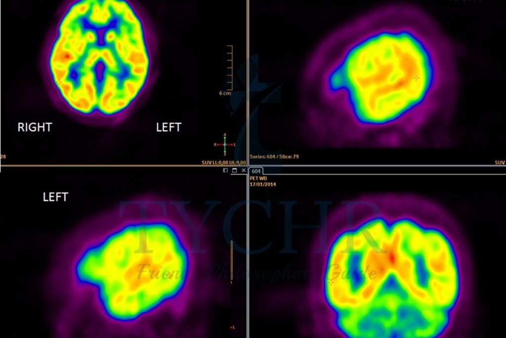
- Positron emission tomography (PET), like fMRI, uses blood flow as the indicator of brain activity.
- A radioactive tracer is used to bind itself to molecules naturally used in the brain, fluorodeoxyglucose {a form of glucose}. This is done into the carotid artery in the neck.. This radioactive tracer is administered into the subject’s blood stream. It has a short period of time (that is, it decays quickly). The frequencies of the radio frequencies emitted by the decaying tracer are registered by the scanner. Brain areas that are more active require more blood supply, so the distribution of the tracer in the brain will depend on what regions are mostly in use at the time of the scan.
- PET has a decent spatial resolution of about 4 mm throughout the brain. However, its temporal resolution is only 30–40 seconds, so quick processes are not easily detected.
- The biggest advantage of PET scans is their good spatial resolution
- They are used less and less these days given the existence of non- invasive alternatives (fMRI) which do not require administration of a radioactive chemical.
- PET is useful for detecting tumors and metastases, as well as other diffuse brain diseases, so that it becomes clear what areas are affected by the spreading disease. It is often helpful in diagnosing causes of dementia.
- Another advantage of PET is that scanners can be small—so small that a small PET scanner has been constructed that can be worn by a rat on its head like a hat. The device is called RatCAP (Schulz et al, 2011). The rat is conscious and fully mobile, it performs various tasks while its brain activity is being measured. This is very useful for research and potentially can lead to a lot of insights into brain functioning.
c) Magnetic Resonance Imaging (MRI)
- MRI’S main principle is the atomic nuclei —in particular those of hydrogen atoms— are able to emit the energy when it’s placed in an external magnetic field. When the scanner detects the pulses of the energy, the relative distribution of hydrogen atoms in the brain can be mapped easily. Hydrogen atoms exist naturally in the body, but their concentration in different types of tissue is different. For example, the highest concentration of hydrogen atoms is found in water (H2O) and fat. Analyzing the pattern of emission of energy in response to magnetic fields, we can see inside the brain.
- After excitation by the magnetic field each tissue returns to its equilibrium state—and the time required to do so differs in different types of tissue. This information is also analyzed. This is why it is necessary to rapidly change the parameters of the magnetic field and switch it on and off repeatedly. The result is the loud noise that is characteristic of any MRI scanner.
The advantages of MRI as compared to CAT include the following.
- It allows non-exposure to radiation and, as a consequence, less risk of radiation-induced cancer.
- MRI has better resolution. This makes it particularly useful for detecting abnormalities in soft tissue—such as the brain.
The disadvantages of MRI as compared to CAT include the following.
- People with metal in their body, for example, cardiac pacemakers or shrapnel, cannot undergo the procedure because metal will attract to the magnetic eld (one can only imagine what happens). Several deaths have been reported in patients with undisclosed metallic implants who underwent the procedure.
- An MRI scan can be an issue for claustrophobic people because it requires being placed in a narrow tube. Also, longer scan times are required: in some cases people have to stay inside the tube for as long as 40 minutes. Specially constructed mirror glasses are sometimes used to create the illusion of openness of the space inside the scanner.
- Lying still for a long time may be problematic for young children, especially since the procedure is new and may be frightening. For this reason, children having MRI scans are often sedated. Some clinics try to turn MRI scans into a fun adventure, pretending that the MRI scanner is a pirate ship, for example.
- An MRI scan is more expensive than a CAT scan. However, the costs are falling.
- Interestingly, the high resolution and sensitivity of an MRI scan is a risk in itself due to incidental findings. Sometimes the scan will then pick up the disturbance or abnormalities in the brain structure which might not be related to the symptoms which can be investigated. This may cause anxiety and cause patients to seek unnecessary treatment.
d) Functional Magnetic Resonance Imaging (FMRI)

- Functional magnetic resonance imaging (fMRI) is called functional for a reason: A dynamic image of the scan is obtained while MRI and CAT are only able to study and show the structural features of the human brain, fMRI can also show the ongoing brain processes.
- In a typical fMRI study the subject is asked to do or carry out a task on which time of the activity is altered with the time of the rest The main principle of this work is that the flow of oxygenated blood increases gradually in that region during the task is performed.. The principle at work is that The response of blood to rapidly changing magnetic fields differs depending on the flow and the level of oxygenation.
- The signal that is analyzed by the fMRI scanner to reconstruct brain activity is known as BOLD (blood-oxygen-level dependent) signal. Other biomarkers also exist but amongst them BOLD is widely used. The energy used by brain cells is directly correlated with the oxygenated blood used by brain cells, and this directly corresponds to the level of the activity in a particular part of the brain..
- An fMRI scan, just like any other brain imaging technique, is characterized by spatial resolution and temporal resolution.
- Spatial resolution is the ability to discriminate between nearby locations: just as with the resolution of your computer screen, the lower it is, the more pixelated the picture and the less detail you can discern. Whereas resolution of your screen is measured in pixels, that of an fMRI scanner is measured in voxels. You can think of them as “volumetric pixels”—a cube of neurons. A voxel is the smallest “brain particle” that we are able to see through a scanner. Typically, The scanner is able to work with the ranges from 1 to 5 mm as the size of the voxel. Small voxels have less blood flow, so the signal is weaker and the required scanning time is longer. A voxel contains several million neurons and several billion synapses. This marks the limit of what can be achieved with brain imaging technology: we can only see a relatively crude picture of brain functioning.
- Temporal resolution is the shortest time period in which the changed brain activity can be registered. Think about the rate as that the snapshots are taken—“frames per second”. Currently, the temporal resolution achieved in fMRI is about 1 second. This also marks a limitation: fMRI is well suited for studying processes that last at least for several seconds (memory, face recognition, thinking about alternatives of a choice and emotional reactions) but is not suited for studying instantaneous processes such as information traveling from the retina to the visual cortex (which takes milliseconds).
- A clear scan requires the subject’s head to be motionless, but this is not realistic. Random thoughts and sensations also result in noise. A lot of noise can be accounted for if the number of trials is sufficient and powerful statistical techniques are used, but some sources of bias are impossible to eliminate.
These are the advantages of fMRI.
- It proposes excellent spatial resolution (up to 1–2mm).
- It grants allowance to us to go through the brain process unlike the usual brain structural imaging technologies, however
There are some disadvantages.
- The resolution of temporal is poor is (about 1 second) comparatively to electromagnetic techniques such as EEG (<1 millisecond) when used.
- All the same things are considered in an MRI and as well as fMRI like the cost of the machine , the claustrophobic claustrophobia, time taken for the procedure and the inability to use it with the implants of medicine.
e) Electroencephalography (EEG)
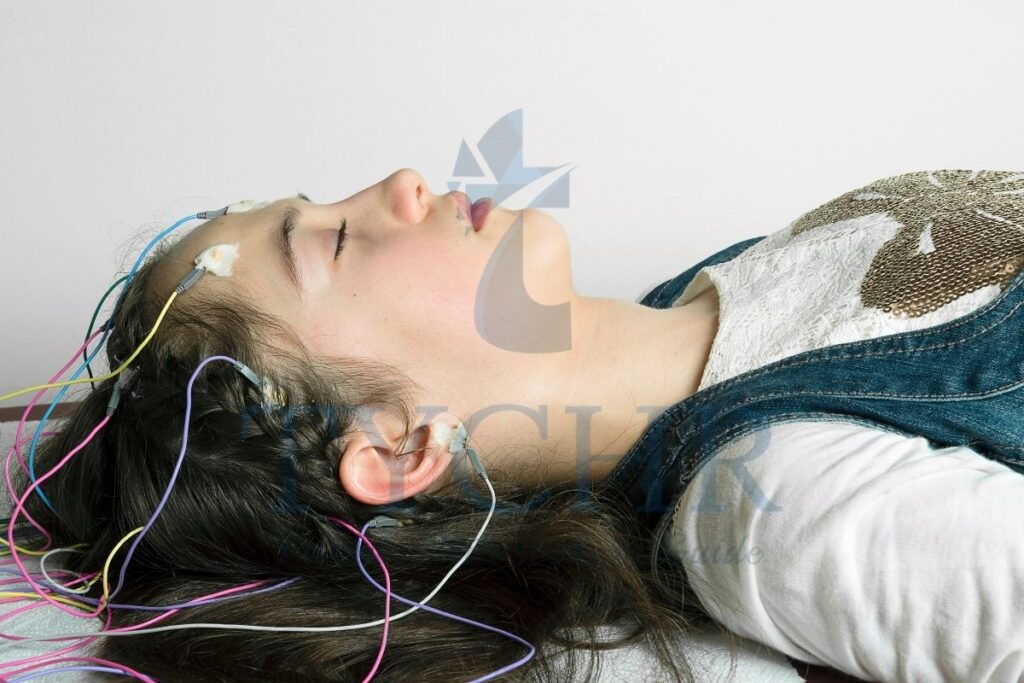
- Electroencephalography (EEG) calculates electric potentials created by neural circuits. Neurons communicate with each other by sending electrical impulses along their axons.
- The impulse Fired in the individual neuron is not seen to any foreign device outside the skull as the impulse is very outside of the skull because the impulse is microscopic. However, when large amounts of neurons fire simultaneously , electric potentials made by these impulses turn detectable at the main surface. Predetermined points on the scalp are inclined toward the electrodes, which detect changes in the electric potential of the scalp regions. This information is vital to generate an electroencephalogram.
- EEG has a perfect temporal resolution. It is capable of detecting changes in brain activity within milliseconds. In this sense it outperforms other techniques such as fMRI.
However , its spatial resolution is a weakness: in practice EEG is never used to set up in the origin of the electrical signal EEG i s good for measuring brain activity “on the whole”. - As it makes visible changes in the overall patterns of brain activity (sometimes referred to as brain waves), EEG is commonly used to diagnose such conditions as epilepsy and sleep disorders.
These are the advantages of EEG.
- It is a low-cost technique.
- Unlike PET and fMRI, EEG measures neuronal activity directly.
- EEG can be offered as a mobile service because the apparatus can be manually transported. For comparison, the weight of an fMRI scanner is about 1 ton.
- EEG is silent, which is an advantage because responses to auditory stimuli can be studied. This is difficult with noisy fMRI scanners.
- EEG is completely non-invasive in comparison to most other neuroimaging techniques.
Using EEG also has disadvantages.
- EEG gives tremendously low spatial resolution, hence it only gives a very ill mannered picture in the means of locality.
- The electroencephalogram (EEG) is designed to measure electrical activity in the cortex, but it is not very good at detecting activity in subcortical areas. Because of the amount of artifacts that contribute to the data’s noise and the low signal-to-noise ratio, correctly interpreting an encephalogram necessitates a great deal of experience. Additionally, the signal becomes weaker the further away it is from the body’s surface. Some possibilities for noise are: heartbeat, muscle movements, eye movements and blinks, and inadequate grounding of the connection between the apparatus and body.
Module 1.7: Animal research role in understanding human behavior
What will you learn in this section
- The role of animal research in understanding behaviour.
- The value of animal models in psychology research.
- Ethical considerations in animal research.
- “Animal research may give an understanding of human behavior”. In some sense this principle is a consequence of the first two principles: “Behavior is the product of physiology” and “Behavior can be genetically inherited”. The physiology and genetic set up of an animal and a human are the same, which naturally proves that to some extent the research of humans is generalizable to animals
The animal’s model value in psychology research.
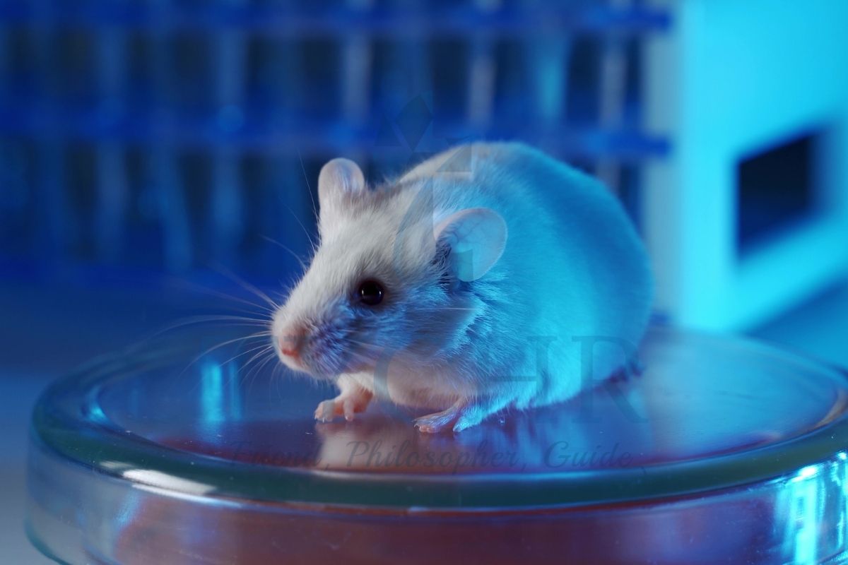
- The amount of animals used in psychological research annually in the USA alone has been estimated to be 1.25–2.5 million, and about 7.5% of psychological research is animal-based (Shapiro, 1998). The most popular species to be used in psychological research are rodents ,dogs, cats, pigeons, hamsters , chimpanzees and baboons.
- The type of Research depends on the purpose of which animal is being used.
1. Researchers in the field of comparative psychology are interested in animal research as an end in itself.They either have an option to focus on a specific species or compare that kind of species to humans. Another group of researchers advocates the study of animals like the models of human beings and the expected result is that the result will be universal in nature and generalizable as well.
2. Human conditions like diseases are researched by the researchers by using animals to understand the particular disease.. - An animal model is a concept that refers to using animal research to test a certain cause–effect hypothesis about a certain human behavior. So an animal model is not just broadly “using animals to understand human behavior”. It is a specific model. For example,There are few animal models which describe depression : stress models (which explain the onset of depression by being more exposed stress triggering situations), separation models (which explain depression by being separated or attachment issues ), medical models (which explain depression cause by the chemical imbalances in the brain), and more.
- There are four types of experimental manipulation tried on animal models (Shapiro, 1998).
- These four types are as follows:
- Invasive manipulations of the nervous system (parts of the brain are stimulated with electrodes, lesion, or removal) and genetic
- manipulation of animals
- Invasive manipulations with different body parts (parts may be restored by substances or damaged)
- behavioral and environmental manipulations (such as electric shocks for rats based on the performance of the rat in the maze).
- Comparison of animal and human brains seemingly conveys a very consistent story of evolution: as species evolved, new structures were built on top of older structures; so the deeper we go into the brain, the more “primitive” structures we will find.
- There is a popular theory of triune brain proposed by MacLean (1990). This theory divides the human brain into three parts:
a)reptilian complex,
b)paleomammalian complex (the limbic system), and
c)neocortex. - The idea is that the deeper brain structures can be found in animals as well; and the further down you go inside the brain, the further down you see in evolution. For example, the reptilian complex that you have in your brain should resemble the full brain of a reptile.
These are some of the advantages.
- Animals and Humans are similar in many ways, specifically in terms of brain structure and genetically.
- Studies with animal models do create results: Because of animal experiment many life saving treatments and useful models of human behavior have been developed gradually. For example, the discovery of insulin as done with the help of experimenting with dogs who had their pancreas removed Several animal studies gives allowance to researchers to work on full life span. While human subjects often outlive researchers themselves, laboratory mice live 2–3 years and this generates as an opportunity to see their nature beyond thor lifespan and even cross generations to generations. This is especially helpful in genetic research.
- Animal research may be highly controlled. For example, the “knockout” technique has been developed to selectively switch off one of the genes in the DNA sequence. All other things being equal, this technique provides great insight into the function of individual genes. The ability to better control confounding variables means higher internal validity of experiments.
- Animal subjects are relatively cheaper and can be easily accessed , easy to manage and manage.
Some of the disadvantages are as follows.
- Animals and humans are not completely similar, and one can never know the extent of the differences between them. This means that animal research, if successful, still needs to be duplicated with humans in order to be sure that findings are generalizable.
- Humans and animals can be similar in some biological aspects but they can completely differ psychologically. (Premack, 2007).
- Researchers typically test new biomedical treatments for mental disorders using mouse models. The results of the rodent experiment, on the other hand, are never directly applied to humans. The drug needs to be tested first on larger animals, even if successful results are obtained using mouse models. It is analogous to a generalization pyramid, with humans at the top and rats at the bottom.
- Since animals are subjected to testing in tightly controlled lab settings, it is possible that they are experiencing stress. As a result, their reactions to experimental manipulations may not be quite the same as in their natural environments: there may be an issue with ecological validity.
- Despite their many similarities, animals and humans are fundamentally distinct. For instance In primates more than 85 antibodies for HIV have worked however it was not something very similar for people.(Bailey, 2008). On the contrary, some results that are negative in animals can actually turn out to be positive in humans. For example, Aspirin was proven dangerous to the animal kingdom but now it’s one of the widely used drugs amongst humans.
Examples of Animal Research
Topic | Study | Findings |
Brain and behavior (localization) | Lashley conducted rodent experiments in which different parts of the vortex were removed to see if memory loss occurred during the maze. | Performance deterioration depends on the percentage of cortex destroyed but not on the location of the destroyed cells. This raises the question against the idea of localizing the memory function. |
Brain and behavior (neuroplasticity) | Merzenich et al (1984): cortical hand representation of the hand in an adult owl. | As the brains’ matter was readjusted, it became in charge of other adjacent digits. |
Ethical Considerations In Animal Research
- Professional associations of psychologists in most countries publish guidelines for research with animals. For example, the American Psychological Association (APA) has a separate document for this purpose. The following document shows the main considered things that should be addressed in all stages of experiment which include animals. Here are just the most crucial of them (APA, 2012)
- Any animal study should be justified “with a clear scienti c purpose”. One of the following justi cations may be used. The study will:
a)increase scientific knowledge of behavior
b)Help us comprehend different species
c)conclusions which are beneficial for human and other animals. - If non-human animals are selected for a particular research its compulsory to ensure that the species which are chosen are the best option to address the research question, and the least number of people required for the non human participants is used, a similar effect on animals.Before conducting the study, all animal research proposals must be submitted to the ethics committee. Psychologists and their assistants must be familiar with the specific characteristics of normal behavior in order to quickly identify animals that are stressed or unhealthy. Lab creatures should be given others conscious consideration. Whenever possible it is advised that the experiments should be designed in such a way that it should cause minimum or no harm to the species. APA guidelines also advise researchers to check with the painful stimuli to be made in use for non-human animals.
- Euthanasia should be performed on any research animal that is observed to be suffering from chronic distress or pain that is not required for the purposes of the thesis study.
- Special cautions are supposed to be taken with the animals who are there in the laboratories; they shouldn’t be let out in the wild.
- Any animal study should be justified “with a clear scienti c purpose”. One of the following justi cations may be used. The study will:
Bateson’s cube: a model used to determine the usefulness of research.
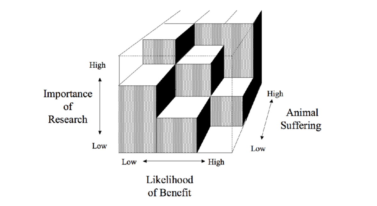
- This model asserts that three domains should he considered:
a)the quality of the research
b)the certainty of medical benefits and
c)the degree of animal suffering. - Research that is of high quality, high benefit medically and low suffering in animals is seen to be worthwhile and has a good purpose. Likewise, poor quality research, with little benefit and high levels of suffering would be seen by this model to be unreasonable.
- Bateson’s model is constructed in such a way that high levels of suffering in animals are unacceptable.
- The cube denotes research that is acceptable [according to this model]. High suffering is never acceptable, but medium suffering in animals, high research quality and high levels of benefit would make the research permissible by these standards.
- Further guiding principles designed to determine the acceptability of animal research are the 3Rs:
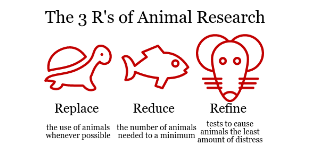
a) Replacement :- Replacement as a principle means that other research methods that replace animal research should be used wherever possible These include the use of cell cultures, human volunteers and computer modeling.
b) Refinement :- Refinement means that research techniques that reduce pain and suffering in animals should be used wherever possible.
c) Reduction :- reduction refers to using research strategies that are efficient and reduce the number of animal experiments used wherever possible.